The shapes and sizes of tissues and organs are critical to multicellular organisms, but the mechanisms controlling the different processes that generate them are only partially understood. In a recent study, a research team, led by Celeste Nelson at Princeton University, linked one of these shape-generating processes, apical constriction, to ATP production by mitochondria. The findings were published in Science Advances.
Patterns of Mitochondrial ATP Predict Tissue Folding
The shapes and sizes of tissues and organs are critical to multicellular organisms, but the mechanisms controlling the different processes that generate them are only partially understood. A research team, led by Celeste Nelson at Princeton University, linked one of these shape-generating processes, apical constriction, to ATP production by mitochondria. The findings were published in Science Advances.
Apical constriction occurs when cells shrink on one surface to fold epithelial tissues. The force is provided by the contraction of actomyosin, and those actions require ATP hydrolysis. Thus, the pattern of ATP hydrolysis is critical. Dr. Nelson and her team sought to determine if the generation of that energy is also spatially patterned.
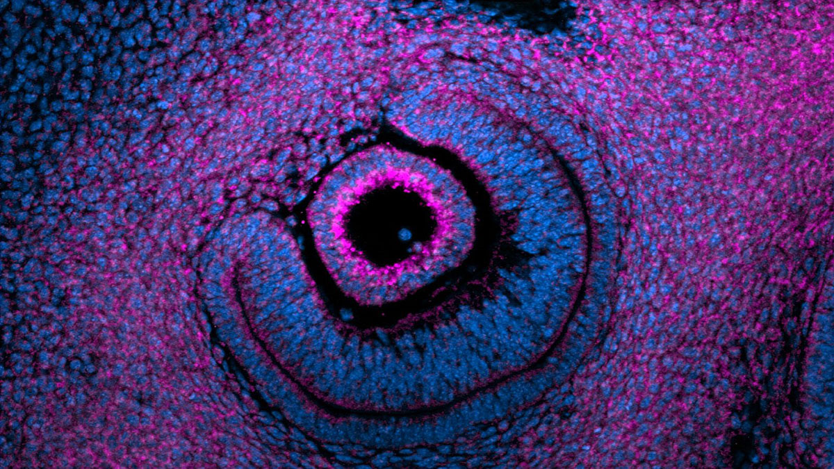
The team examined ventral furrow formation during gastrulation in Drosophila, neural tube closure during neurulation in chicken, branch formation during lung development in chicken, and lens placode invagination during eye development in mouse embryos. During development, these tissues use apical constriction to fold the epithelium in a well-characterized pattern. The researchers determined mitochondrial density by immunofluorescence staining for Tom20, an outer mitochondrial membrane protein. Using that and other tools (e.g., time-lapse imaging, spatial transcriptomics, oxygen consumption rate), they found that mitochondrial density, membrane potential, and ATP production are greater on the apical sides of epithelial cells during apical constriction. In fact, mitochondrial density can be used to predict which cells will undergo apical constriction before they initiate the fold.
During development, tissue shape depends on multiple factors, including gene expression, mechanical forces, and energy. These processes are remarkably conserved. The Nelson team demonstrated that the location of mitochondria predicts apical constriction and is upstream of other activities. Their findings implicate the spatial distribution of bioenergetics in development.
A Statement of Significance by Dr. Nelson
Many developing tissues generate their final shapes through a series of highly reproducible and stereotyped folds, akin to origami patterns. Over the past few decades, the field has uncovered many of the gene expression changes and biochemical signals that are necessary to generate the forces to fold these tissues. Our work now shows that ATP is generated at the site of folding before it begins by oxidative phosphorylation, and that mitochondria concentrate at these sites. So now we need to figure out whether the same genetic and biochemical signals are leading to both mitochondrial localization as well as force induction, or whether there are parallel signals.
A Conversation with Dr. Nelson
MitoWorld: Can you give us an idea of what direction your research might take to further your findings here?
Dr. Nelson: There is an increasing recognition in the field that energy metabolism and mechanical forces are interacting with each other on some scale within cells and tissues, and we’re seeing more and more research groups exploring these interactions, which is very exciting! Our group is primarily focused on branching epithelia, so we’re currently exploring the different ways in which epithelial branching morphogenesis is fueled across systems, whether it’s primarily by mitochondrial oxidative phosphorylation, or by glycolysis, or both.
MitoWorld: You focused on apical constriction in this work. Do you think the findings here can be extrapolated to other mechanisms involved in tissue morphology?
Dr. Nelson: Our data suggest that there’s an interesting coupling between energy metabolism and tissue folding. Since we published our findings, several other scientists have mentioned that they’ve also observed mitochondria at the apical side of their apically constricting tissues! So we think this observation might represent a conserved energetic-mechanical motif. But as your question suggests, tissues can fold in ways that don’t involve apical constriction. It makes sense to assume that other folding mechanisms require energy metabolism, but I wouldn’t be so bold as to predict that spatially patterned mitochondria are universal across types of folding events.
MitoWorld: Can you speculate on how mitochondria are controlled by the other elements of the signaling apparatus that determines tissue morphology? Your last sentence in the Discussion suggests the complexity of this question.
Dr. Nelson: The most stunning observation that we made, in my opinion, was that mitochondria appear at sites where energy is needed before the tissue folds. How do they know to get there? What moves them there? I wish that I could say we know the answer to those questions, but we don’t. The fact that we observe this patterning across species and systems suggests something fundamental to me. It’s probably not dependent on a specific growth factor or morphogen, but rather coupled to the subcellular changes necessary for a block-like epithelial cell to change shape in the first place.
MitoWorld: This is somewhat related to the previous question. Do you have any idea of how the mitochondria are held in position at the apical side of the cell?
Dr. Nelson: I’m not sure they’re actually held there, per se. We have some timelapse imaging data that suggest the mitochondria are fairly dynamic, moving along the apical-basal axis of the cell. It would be really cool to do a pulse-chase experiment with mitochondria to see how many persist on the apical side and how many move back and forth.
MitoWorld: You note an interesting balance between glycolysis and oxidative phosphorylation. Can you elaborate on that relationship?
Dr. Nelson: In many systems, it’s been reported that cells use either glycolysis or oxidative phosphorylation (oxphos) in lieu of the other. We don’t see that to be true for apical constriction, at least for the developing chicken lung where we conducted seahorse experiments. In that system, we found both increased oxphos and increased glycolysis in the regions of tissue undergoing apical constriction. So there’s certainly more to be learned about the energetics of apical constriction, and about how the cell budgets energy generated by oxphos versus glycolysis.
Reference
Lemma B, Rothstein M, Zhang P, Waas B, Kilwein M, Topiwala S, Zhang SX, Sudhakar A, Goodwin K, Gavis ER, Mallarino R, Kosmrlj A, Nelson CM (2026) Patterns of mitochondrial ATP predict tissue folding. Science Advances 12(17): eaee6175.