
Mitochondria are widely known for being the major site of ATP production in cells. The two enzyme pathways are oxidative phosphorylation (OXPHOS) and the tricarboxylic acid (TCA) cycle. The only structural link between these two systems was thought to be succinate dehydrogenase (complex II).
A research team, led by Nils-Göran Larsson (Karolinska Institute) and Albert J. R. Heck (Utrecht University), wondered if there might be other connections. They used multiple procedures, including in-solution crosslinking mass spectrometry, to determine the location of ATP synthase in multiple mouse models and tissues. They found that ATP synthase (complex V) interacts with several TCA enzymes. The results were published in the journal Nature Communications.
The researchers found that portions of ATP synthase interact with the TCA cycle enzymes. They also showed that disrupting mitochondrial DNA gene expression destabilizes ATP synthase and induces additional interactions with the TCA cycle. They also took advantage of heart- and skeletal-muscle-specific Lrpprc-knockout mice. Using this well-established model of impaired mitochondrial gene expression, the team found that ATP synthase was remodeled and increased the interactions with various metabolic proteins in the mitochondrial matrix.
The study showed that ATP synthase is remodeled in response to impaired gene expression. This work documents how mitochondria may adapt their metabolism to accommodate the loss of key mitochondrial enzymes.
A Statement of Significance by Drs. Larsson and Misic
Our study adds a new dimension to our understanding of mitochondrial energy metabolism by revealing previously unrecognized spatial connections between ATP synthase and several TCA-cycle enzymes. These findings suggest that the molecular organization of mitochondrial energy metabolism is more interconnected than previously appreciated. Particularly intriguing is the observation that these interactions become more prominent when mitochondrial gene expression is impaired, suggesting that the physical organization of metabolic enzymes is dynamic and undergoes remodeling during mitochondrial dysfunction. Although the physiological significance of these interactions remains to be established, they raise important questions about whether such rearrangements contribute to metabolic adaptation under conditions of impaired energy production. More broadly, our findings provide a new perspective on how mitochondrial metabolic machinery is organized and remodeled, opening new avenues for understanding mitochondrial function in health and disease.
A Conversation with the Authors
MitoWorld: Can you give us an idea of what directions your research might take to further advance the findings of the current paper?
Drs. Larsson and Misic: We have a long-standing interest in the organization of the respiratory chain in physiology and disease. Why do the respiratory chain complexes from supercomplexes? We previously found that the supercomplexes do not increase the efficiency of the respiratory chain or prevent ROS production. We now report unexpected novel connections between the ATP synthase and TCA cycle enzymes.
MitoWorld: Your results show that ATP synthase interacts in complex ways with multiple TCA enzymes that are affected by mtDNA mutations. Are these many interactions simply to facilitate energy production or is there another rationale?
Drs. Larsson and Misic: The physiological importance of the interactions between the ATP synthase and the TCA cycle enzymes is unknown. These interactions increase when mtDNA gene expression is impaired and may provide a compensatory mechanism.
MitoWorld: The ability of the multi-enzyme complexes to adapt to different metabolic demands is amazing. Can you speculate on the mechanism that allows the interactions to rearrange themselves under these circumstances?
Drs. Larsson and Misic: Our data show a protein-protein interaction between the ATP synthase and some TCA cycle enzymes, such as malate dehydrogenase (MDH). Interestingly, a recent Cell paper on proteomics of microorganisms has shown that Acanthamoeba has a stable interaction between ATP synthase and MDH, arguing that this interaction may be important for metabolism.
MitoWorld: XL-MS is a powerful tool for exploring protein interactions. Do you plan to use it to examine other aspects of OXPHOS?
Drs. Larsson and Misic: We already used it to study the organization of respiratory chain supercomplexes in the mitochondrial inner membrane.
MitoWorld: Do your findings have any potential clinical applications, and if so, do you have any plans to follow up on those?
Drs. Larsson and Misic: Our study is a basic science contribution and much more work is needed to determine if the interactions we report can provide a target for drug development.
Reference
Pérez Pañeda L, Misic J, Kadavá T, Larsson NG, Heck AJ (2026) Investigating the relationship between ATP synthase and the TCA cycle by crosslinking mass spectrometry. Nature Communications 17(1): 5563. https://www.nature.com/articles/s41467-026-74730-5.pdf
Maintaining body temperature is a critical requirement for all mammals. Multiple circadian, environmental, and dietary factors are involved in regulating the mitochondria in adipose cells to produce heat. An international research team, led by Zachary Gerhart-Hines of the University of Copenhagen, found that all of these factors converge on the mitochondrial metabolite transporter SLC25A34. The findings were recently published in the journal Science.
The production of heat by thermogenic adipose cells is regulated by circadian rhythms, but environmental factors, physical activity, and other factors can override the circadian control to more precisely match lipid metabolism to the immediate need. Interestingly, lipid metabolism in the brown adipose tissue can increase lipid synthesis in the cytosol and fatty acid b-oxidation in the mitochondria at the same time. How the body deals with the circadian rhythms and responses to acute cold are not understood.
The research team sought to clarify this complex regulation. They noted that Slc25a34 mRNA was expressed in a daily rhythm. During sleep the nuclear receptors REV-ERBα and REV-ERBβ cause histone deacetylase 3 to shut off expression of Slc25a34 mRNA. Upon waking, the repression is ended, and expression resumes. Other factors, such as cold or a lipid-rich meal, increase expression. The researchers used biochemical and metabolic tests to show that levels of SLC25A34 increase and decrease according to the body’s needs. The model they developed depends on the cycle of synthesis of lipids in the cytosol, and the oxidation of fatty acids in the mitochondria.
Their findings show that heat production by thermogenic fat tissue is controlled by multiple factors, but they all come together in the protein SLC25A34. This model greatly expands our understanding of this key physiological activity.
Statement of Significance by Dr. Gerhart-Hines
Fat cells have to reconcile two very different kinds of information: the predictable rhythm of the day and unpredictable changes in temperature and diet. We found that all three inputs converge on a single, previously uncharacterized mitochondrial carrier, SLC25A34. During sleep the clock holds it off through the REV-ERB repressors and HDAC3; waking, cold, and lipid-rich diets release that brake and switch the gene on through PPARα and PPARγ. Our data indicate that SLC25A34 brings oxaloacetate back into mitochondria, which keeps the TCA cycle turning and sustains the cytosolic acetyl-CoA needed to build new lipids. That is what allows thermogenic fat to make and burn lipid at the same time, an energy-dissipating cycle that has been recognized for years but whose timing and control were not understood.
A Conversation with Dr. Gerhart-Hines
MitoWorld: This study was an amazing team effort. Can you give us an idea of how you plan to further this work in the future?
Dr. Gerhart-Hines: Our first priority is to address direct transport at the biochemical level. Our metabolomics, compartment-resolved measurements, isotope tracing, and molecular docking all point to oxaloacetate import, but the definitive experiment is to reconstitute the purified carrier in liposomes and measure transport directly. That will also tell us whether N-acetylaspartate, malate, or other dicarboxylates are part of the picture. The second priority is to look beyond brown fat. At thermoneutrality, the heart has the highest Slc25a34 expression of any tissue we examined, followed by slow-twitch muscle, kidney, and several brain regions, and the REV-ERBs and PPARs that control it are active in all of them. Our third goal is to expand on understanding the human relevance of the transporter. Knocking down SLC25A34 in brown adipocytes from four donors reduced their respiratory capacity, and in subcutaneous white fat, its expression tracks with a healthier metabolic profile across 24 clinical studies. We now want to find out whether that relationship is causal.
MitoWorld: Body temperature is critical to mammals, and the regulation involved in these activities is exquisite with multiple players. SLC25A34 is one example. What is the next most-important question to resolve?
Dr. Gerhart-Hines: For SLC25A34, the question I most want answered is why it exists at all. Cells already have the malate-aspartate shuttle to move these carbon backbones between compartments, yet thermogenic fat switches on a separate, tightly regulated carrier that appears to run in parallel. Our working idea is that every time citrate or N-acetylaspartate is used to make cytosolic acetyl-CoA, an oxaloacetate is left behind in the cytosol, and SLC25A34 returns it to the matrix so the cycle can keep running at high flux. Proving that means defining what the carrier directly transports and in exchange for what. A related puzzle is its paralogue, SLC25A35. Both descend from the same yeast oxaloacetate carrier, but SLC25A35 is high at baseline in brown fat, falls in the cold, is not under clock control, and knocking it down did not affect respiration in our hands. Shingo Kajimura’s group recently linked it to phosphoenolpyruvate export for glycerolipid synthesis. Why mammals kept two versions with such different regulation is something I would love to understand.
MitoWorld: The final paragraph of the Discussion includes some intriguing speculations. For example, can you expand on the potential role of SLC25A34 in the brain?
Dr. Gerhart-Hines: I should stress first that this is speculation, since we have not done any experiments in the brain. The idea comes from N-acetylaspartate (NAA). When we depleted SLC25A34 in brown adipocytes, NAA was the most strongly changed metabolite, even after a single day, and the pattern fits NAA becoming trapped in the mitochondria, where it is made. NAA is best known from the nervous system. It is the second most abundant metabolite in the brain and supports myelination and neuronal integrity, and yet, how it leaves mitochondria has never been resolved. Slc25a34 is expressed in several brain regions, and so, it is reasonable to ask whether the carrier contributes to NAA handling there as it appears to in fat, either directly as a counter-exchange partner for oxaloacetate or indirectly. Testing that in neurons is an obvious next experiment, and one where we would be glad to receive some collaborative help from the neuroscience community.
MitoWorld: Might your findings have any clinical implications? If so, what might be the “low-hanging fruit”?
Dr. Gerhart-Hines: We need to be careful here because this was a mechanistic study done primarily in mice and cultured mouse and human cells. That said, the lipid cycle that SLC25A34 supports burns energy by design, and increasing that kind of cycling is one of the strategies being considered to counter the caloric excess of obesity by numerous pharmaceutical companies. The human data are encouraging: the carrier is needed for full respiratory capacity in human brown adipocytes, and higher expression in subcutaneous fat goes along with lower BMI, triglycerides, LDL cholesterol, and insulin resistance. Those are correlations, not proof. I don’t think the low-hanging fruit is a drug aimed at the carrier itself. Activating a transporter is hard, and with expression so high in the heart, we need to understand what it does there first. The nearer-term opportunities are to use SLC25A34 as a readout of lipid-cycling capacity in human fat, and to build on what we learned about its regulation. It is one of the most selective PPARα targets we found in brown fat, and it responds to time of day, cold, fasting, and dietary fat, so we can now ask when and how existing interventions engage this pathway.
MitoWorld: This paper has 65 authors. Can you give us an idea of how you organized such a large number of researchers at multiple institutions and in several countries?
Dr. Gerhart-Hines: This comprehensive project developed gradually from one question at a time over more than a decade. The seeds of the project were already planted while I was a postdoc with Mitch Lazar at the University of Pennsylvania. When I came to Copenhagen to start my lab in 2014 at the Novo Nordisk Foundation Center for Basic Metabolic Research, this initial discovery formed the basis of my first grant as an independent investigator. Iuliia Karavaeva, the first author, carried it for the better part of those years and was the one person who held the whole story in her head. Each time the data raised a question we could not answer ourselves, whether that was measuring a metabolite as unstable as oxaloacetate in separate cell compartments, isotope tracing, carrier structure, human brown adipocytes, or a particular mouse model, we went to the people who were experts in that area. That is how we ended up with colleagues in eight countries. What made it work was each collaborator’s passion for the science and their generosity in staying with us through the long haul. Being able to work with so many gifted and brilliant experts across disciplines was one of the highlights of this project for me.
MitoWorld: Mitochondria are involved in so many key cellular activities. How did you come to be interested in them?
Dr. Gerhart-Hines: I have been fascinated by mitochondrial biology since my PhD studies in Pere Puigserver’s group at the Dana-Farber Cancer Institute, where I worked on the sirtuins in muscle. Later, as a postdoc with Mitch Lazar, I studied how the circadian repressor REV-ERBα controls brown fat thermogenesis. When we went looking for factors controlled by both the molecular clock and cold, only two met every criterion: UCP1, the classic thermogenic protein, and an orphan carrier nobody had studied, SLC25A34. Both sit in the inner mitochondrial membrane and belong to the same SLC25 family. Once my work on the clock and fat tissue had converged on this uncharacterized mitochondrial transporter, I leapt at the opportunity to bring together many of my scientific interests in adipose metabolism, mitochondrial biology, and energy homeostasis. The result also made it clear to me that the decisions a fat cell makes about storing or burning energy are heavily shaped by what crosses that membrane. The project also highlighted how mitochondria play roles far beyond acting as the so-called ‘powerhouses of the cell’. Through metabolites like acetyl-CoA they also report back to the nucleus, and this carrier turned out to sit right at that interface.
Reference
Mitochondria are involved in multiple activities within cells, including energy, biosynthesis, redox balance, and apoptosis. Furthermore, mitochondrial DNA (mtDNA) mutations are common in cancers, but how they affect tumor growth and the response to therapies is not well understood. A recent review, by Laura Greaves and colleagues, explored the prevalence and effects of mtDNA mutations in cancer, how they affect metabolic plasticity, and new experimental models and potential therapeutic opportunities. The study was published in Trends in Molecular Medicine.
The review emphasizes several key points. mtDNA mutations are common across human cancers. Mutation rates vary by tissue but range from 13% in glioblastoma multiforme to 63% in rectal adenocarcinomas. The effects of the mutations depend on the degree of heteroplasmy. Complex 1 is particularly affected, and there seem to be upper and lower limits to the levels of heteroplasmy that affect tumor progression. One common deletion (i.e., nucleotides 8470–13,447) that eliminates five tRNA genes and seven OXPHOS subunit genes is selected against during tumorogenesis. These findings suggest that the deletion is detrimental to cancer cell proliferation.
Recently, several exciting new mtDNA mutation experimental systems have been developed. For example, the transmitochondrial cytoplasmic hybrids involve the fusion of a mtDNA-depleted nuclear donor and an enucleated cell with mtDNA with specific mitochondrial genotypes. The mitochondrial genome can now be directly edited with DddA-derived cytosine base editors that enable precise C:G-to-T:A conversions within mtDNA. The PolgA mouse model allows mtDNA mutations to accumulate much faster than before. A point mutation reduces the effectiveness of proofreading by DNA polymerase gamma.
Yet, several significant challenges remain. It is difficult to interpret studies of mtDNA in tumors. Bulk homogenates contain the DNA of malignant cells, making it hard to know the exact amounts of DNAs in the mass. Even the hybrid models have limitations.
This comprehensive review provides an excellent summary of mitochondria and mtDNA in cancer. It summarizes the current understanding of these mutations in tumors and the opportunities and challenges of future research. Recognizing the effects of mtDNA mutations on mitochondrial metabolic plasticity and intercellular mitochondrial transfer may suggest new therapeutic approaches.
A Statement of Significance from Dr. Greaves
In this review, we bring together emerging evidence on how mitochondrial DNA mutations influence cancer metabolism, immune interactions and treatment response. We highlight that their consequences depend on the specific mutation, heteroplasmy level and cellular context, and explain how advances in mitochondrial genome editing now enable direct tests of causality. By connecting these experimental opportunities to therapeutic questions, we identify priorities for determining when mtDNA mutations create vulnerabilities that could inform more effective cancer treatments.
A Conversation with Dr. Greaves
MitoWorld: This is a terrific overview of mtDNA mutations and their involvement in cancer. Have you had any reaction from the research community as yet?
Dr. Greaves: It’s only recently been published, so it’s still early to gauge the response from the research community. It has already been cited, which is encouraging! I’m looking forward to seeing the conversations and research it helps to stimulate.
What I hope comes across is that we need to understand these mutations in context. Finding an mtDNA mutation in a tumour does not, by itself, tell us what it is doing. Its effects may depend on the particular mutation, its abundance and the biology of the tumour. The exciting thing is that we now have tools to investigate those questions experimentally, including whether the adaptations these mutations provoke could provide opportunities for treatment.
MitoWorld: Can you describe how your own research fits into the topics covered in this review?
Dr. Greaves: My research has grown out of an interest in how mtDNA mutations accumulate as we age and what they do to cells, particularly in the intestine. That led naturally to questions about cancer: how might these mutations affect tumour development, and could they influence how a tumour responds to treatment?
We are now investigating those questions using complementary approaches in colorectal, gastro-oesophageal and ovarian cancers. Working with our collaborators, we have introduced specific mtDNA mutations into organoids and human cancer cell lines, which we can compare with matched isogenic controls. That allows us to move from observing an association to testing whether a mutation actually causes a change in tumour behaviour.
We want to understand how these mutations affect growth, metastasis and interactions with surrounding cells, and whether the metabolic adaptations they produce create vulnerabilities we can target. Connecting that experimental work back to human tumours is central to what we are trying to achieve.
MitoWorld: Heteroplasmy is a significant challenge for studying the effects of mtDNA mutations and many other aspects of mitochondrial biology. The model systems seem to be a promising strategy to overcome this “threshold” challenge. Can you expand on those challenges and possibilities?
Dr. Greaves: The complication is that a cell contains many copies of mtDNA. Some can carry a mutation while others do not; that mixture is what we call heteroplasmy. The remaining normal copies can compensate, so a mutation may need to reach a substantial proportion before we detect a respiratory defect. There is no single threshold that applies to every mutation or cell type.
An average measurement across a tumour can also hide very different situations. It could reflect similar mutation levels in every cell, or a mixture of cells with very high and very low levels. Those patterns can change over time. Base editing and single-cell measurements give us ways to investigate that complexity directly.
For our models, the opportunity is to compare cells carrying the same mutation at different measured heteroplasmy levels and follow both the mutation and its functional effects. We cannot simply set any percentage we want and assume it will remain stable. These models allow us to study the threshold itself—and ask whether the threshold for altered growth or drug sensitivity is the same as the threshold for impaired respiration.
MitoWorld: The migration of mitochondria from one cell to another must be another challenge to the study of mtDNA mutations. Do you see any light at the end of the tunnel?
Dr. Greaves: Yes, although it certainly adds another layer of complexity! We have to consider the possibility that a cell’s mitochondrial population can be influenced by its neighbours. There is evidence in experimental cancer systems that the transfer of mitochondria carrying mtDNA mutations from cancer cells into T cells can impair their function. That makes transfer a potentially important part of tumour biology, as well as something we need to account for experimentally.
I think the way forward is to combine imaging with genetic tracking and functional measurements. We need to establish where the mitochondria came from, whether their DNA persists in the recipient cell and whether the transfer changes what that cell can do. Seeing mitochondrial material move is only part of the story.
The questions I would want to ask are how frequently this happens in tumours, under which conditions, and whether it helps cancer cells survive or changes the immune response. Answering those questions could reveal another way to interfere with the support that tumours receive from their surroundings.
MitoWorld: The shared governance of the cell between the nuclear genome and the much smaller mitochondrial genome is a fascinating topic. The mtDNA mutations present another aspect of that governance. Can you elaborate on how the study of these mutations might be window on the interactions of the two genomes?
Dr. Greaves: The two genomes have to work together for mitochondria to function. MtDNA encodes just 13 proteins, all involved in oxidative phosphorylation, while the nuclear genome supplies the vast majority of mitochondrial proteins. A change in either genome therefore has to be understood in the context of the other.
This is a central question in our research programme. If we introduce the same mtDNA mutation into cells with different cancer-driving nuclear mutations, do those cells respond in the same way? Can some compensate more effectively, and does that compensation create a particular drug sensitivity?
We can then test the explanation by manipulating candidate nuclear genes and asking whether that removes or restores the phenotype. That takes us beyond describing differences and towards identifying the mechanisms responsible. Ultimately, I want to know whether understanding both genomes together will help us predict tumour behaviour and treatment response more accurately.
MitoWorld: How did you come to be interested in mitochondria in the first place?
Dr. Greaves: It was quite an unexpected route. I studied pharmacology at Newcastle, and my undergraduate research project was the point at which I realised how much I enjoyed research. I liked being able to investigate a question where the answer was not already known.
I then started a PhD with Doug Turnbull and Tom Kirkwood, initially intending to study mtDNA mutations in neuronal stem cell ageing. When I arrived, though, the group had just received some colon tissue, and I was asked to look at mitochondrial function in the colonic epithelium before starting the planned project.
That ended up becoming my PhD and setting the direction of my research. I never got to the neurons! Instead, that initial work in the colon developed into a lasting interest in mtDNA mutations, ageing and, eventually, cancer. It is a good example of how an unexpected observation can shape an entire research career!
Reference
Shang E, Aftab O, Dayanamby A, Greaves LC (2026) Cancer mtDNA mutations: Metabolic plasticity and therapeutic promise? Trends in Molecular Medicine Cancer mtDNA mutations: metabolic plasticity and therapeutic promise?: Trends in Molecular Medicine
For some time, tau has been an enigmatic protein. Aggregations of tau form the tangles reported by Alois Alzheimer as one of the features of Alzheimer’s disease. Tau is thought to stabilize microtubules and participate in other functions in brain neurons. However, pathological forms of tau (e.g., hyperphosphorylated tau, aggregations of tau) are associated with tauopathies, such as Alzheimer’s disease, frontotemporal dementia, progressive supranuclear palsy, and Pick’s disease. Yet, the link between tau and those diseases is unclear.
The team led by Dr. Lu sought to better understand that link. The results of their study were recently published in the journal Neuron.
The team used three experimental systems: flies, mice, and human iPSCs carrying a mutation that causes tau hyperphosphorylation and induced them to form neurons. By manipulating these cells, they found that reverse electron transport (RET) was enhanced. The result is that the cells produce more reactive oxygen species and have lower NAD+/NADH ratios. The researchers found that tau enters the mitochondria and interacts with complex 1 subunit NDUFS3. These actions cause increased RET and tau hyperphosphorylation.
The key finding of the study was to show that tau regulates RET, and that interfering with this this normal function can cause disease. It also found that inhibiting RET reduces tau levels and the risk of tau toxicity. Thus, regulating RET might be a promising therapeutic target.
A Statement of Significance by Dr. Lu
A major hurdle to understanding the core pathogenic mechanisms of tau and developing effective therapies is the uncertainty of the normal physiological function of tau that becomes altered in disease. This study provides across species evidence that tau plays a physiological role in facilitating mitochondrial reverse electron transport (RET), presumably as a stress adaptation mechanism. However, persistent RET activation by tau mutations, chronic stress, or aging becomes pathological, driven by a self-perpetuating vicious cycle in which phosphorylated tau (p-tau) promotes RET, and RET further promotes tau phosphorylation through tau kinases. Pharmacological inhibition of RET with a brain penetrant small molecule breaks the vicious cycle and offers therapeutic benefits in animal models and human iPSC derived neuronal models, nominating RET inhibition as a viable therapeutic approach for a broad spectrum of brain diseases that involve tau.
A Conversation with Dr. Lu
MitoWorld: Can you give us an idea of the direction you might take to advance the research contained in this paper?
Dr. Lu: This study opens many interesting research directions: 1) What is the normal physiological function of reverse electron transport (RET)? We know it is activated by stresses such as higher ambient temperature. But we know little about its physiological role in stress adaptation and response. 2) Other than tau, what other cellular proteins can modulate RET? 3) At the structural level, how does tau binding to mitochondrial complex I promotes RET, and how can this be inhibited by small molecule drug candidates? 4) How are the reverse and forward electron transport processes coordinated and differentially regulated? These are the research directions we are actively pursuing.
MitoWorld: Your work shows interaction with several complex systems (e.g., sirtuins, NAD+, ROS, post-translational modifications, protein aggregations). The inability to unravel all of these has thwarted research into Alzheimer’s disease for example. Can you speculate how these might fit together?
Dr. Lu: It is possible that the seemingly disparate, complex systems mentioned above may not be so isolated from each other after all. The fact that RET inhibition ameliorates disease phenotypes and modifies all these systems suggests that RET activation may sit at the top of the cascade and drives these different aspects of disease phenotypes. For example, RET generated ROS can activate tau kinases to promote tau hyperphosphorylation and aggregation. RET induced NAD+/NADH ratio change can alter cellular metabolism and impact the activities of signaling molecules such as the Sirtuins, which use NAD+ as co-factors. Previous efforts have targeted the downstream effector of RET individually, explaining their limited clinical success in Alzheimer’s disease and other neurodegenerative diseases.
MitoWorld: You mention that regulating RET might have clinical implications. Can you speculate on how that might be done, and do you have any plans to pursue such studies?
Dr. Lu: Our study indicates that RET acts early in the disease process and that RET and p-tau forms a self-perpetuating pathological loop that drives tauopathies. Our results also show that CPT, a small molecule that inhibits p-tau binding to mitochondrial complex I, breaks the RET/p-tau pathological loop and offers therapeutic benefits in multiple animal models and human iPSC-derived neuronal models of tauopathies. CPT belongs to Cerepeut, Inc, which is currently conducting IND-enabling studies on the compound.
MitoWorld: Can you tell us how you came to be interested in mitochondria and their roles in physiology and disease?
Dr. Lu: I first became interested in mitochondria when I was an undergraduate student at Fudan University where I did my thesis research on plant mitochondria. I was fascinated by mitochondria for their origin: The evolutionary origin of mitochondria is explained by the endosymbiotic theory, which suggests that mitochondria were once free-living prokaryotes that entered a symbiotic relationship with a host cell some billion years ago. This transformation from an independent organism to a specialized organelle is perhaps one of the most significant events in the history of life, because it enabled the development of complex multicellular organisms. Mitochondria are also fascinating because of their versatile roles in biology: They are best known as the power plant of cells due to their fundamental roles in making ATP, the currency of energy. But they do more than just making energy. It is becoming increasing clear that they are critical signaling organelles that control the proliferation, differentiation, and survival of the host cells. As a result, dysfunction of this organelle features prominently in diverse human diseases. Mitochondria are truly iconic organelles in biology and medicine.
My scientific career is intimately intertwined with mitochondria. I did my PhD at Cornall University, where I worked with Dr. Maureen Hanson on mitochondrial biology, specifically, RNA editing in plant mitochondria. For my postdoctoral training I wanted to do something different. I joined the Jan lab at UCSF, where I changed my research direction to neuroscience, studying the behavior of neural stem cells in fruit flies. After my postdoc, I established my independent lab first at the Rockfeller University in New York city and later at Stanford University, where in addition to continuing the work initiated during my postdoc, I started new research directions, working on the mechanism of brain diseases, from neurodegenerative diseases to brain tumor. It is the disease related research that led me to bump into mitochondria again. For example, our work on PINK1 and Parkin, two genes linked to familial Parkinson’s disease, and our work on the Notch signaling pathway in cancer biology have all implicated the fundamental involvement of mitochondria.
By Chiara Zurzolo and Ines Saenz-de-Santa-Maria
Mitochondria can be exchanged between cancer cells and cells of the tumor microenvironment, but the mechanisms and functional consequences of this exchange remain incompletely understood. Recently, Ines Saenz-de-Santa-Maria, a former postdoctoral researcher in Chiara Zurzolo’s laboratory at the Institut Pasteur and currently a postdoctoral researcher at CIBERONC, supported by the ARISTOS programme, and Zurzolo led a study investigating how tunneling nanotubes (TNTs) mediate mitochondrial exchange between glioblastoma cells and non-tumoral astrocytes.
The study reveals a potential mechanism by which intercellular mitochondrial exchange may contribute to tumor adaptation and highlights TNT-mediated communication as a potential therapeutic target. The results were published in Nature Communications.
Mitochondrial transfer between tumor cells and between tumor and non-tumor cells has been implicated in tumor progression, metabolic adaptation and resistance to therapy. One important route for this transfer is through TNTs, thin membranous connections that establish direct communication between cells. TNTs are heterogeneous in their cytoskeletal composition: some contain predominantly actin, whereas others contain both actin and microtubules and tend to be thicker and more stable. In glioblastoma, TNTs coexist with another type of intercellular connection known as tumor microtubes, creating complex networks of communication within the tumor and its microenvironment.
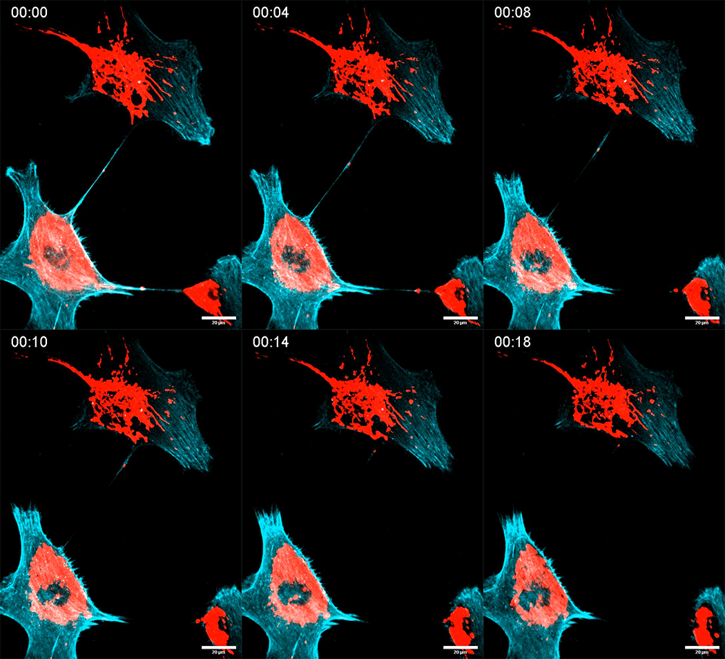
Mitochondrial transfer between tumor cells.
Saenz-de-Santa-Maria and colleagues investigated mitochondrial transfer between glioblastoma/glioblastoma stem cells and non-tumoral astrocytes. They found that mitochondrial exchange is bidirectional but asymmetric, with astrocytes transferring mitochondria to tumor cells considerably more efficiently than tumor cells transfer mitochondria to astrocytes. Importantly, the consequences of transfer depended on its direction. Astrocyte-derived mitochondria integrated into the mitochondrial network of recipient tumor cells, and their acquisition was associated with changes in mitochondrial activity and increased expression of genes involved in mitochondrial metabolism. Conversely, damaged mitochondria transferred from glioblastoma cells to astrocytes were targeted to the lysosomal compartment and underwent mitophagy, suggesting that astrocytes may contribute to the disposal of dysfunctional tumor-cell mitochondria.
The researchers also asked whether TNT-mediated mitochondrial communication occurs in tumors in vivo. Using intravital subcellular microscopy in living mice, they visualized TNT-like connections between tumor cells with characteristics similar to those observed in vitro, including mitochondria within these structures. They also detected transferred mitochondria in recipient cells. These observations support the existence of TNT-mediated mitochondrial transfer in living tumors, although the complexity of the tumor environment makes it difficult to completely distinguish TNTs from other intercellular structures such as tumor microtubes.
The findings reveal a bidirectional mitochondrial exchange between tumor cells and their microenvironment that contributes to mitochondrial homeostasis. Astrocytes can provide tumor cells with healthy mitochondria associated with metabolic changes, while at the same time accepting damaged tumor-cell mitochondria that can subsequently undergo mitophagy. The results, therefore, suggest that intercellular mitochondrial exchange may constitute a previously underappreciated mechanism of tumor adaptation and resilience and raise the possibility that disrupting TNT-mediated communication could provide a new therapeutic strategy.
A Statement of Significance by Chiara Zurzolo and Ines Saenz-de-Santa-Maria
Our study reveals that mitochondrial exchange between glioblastoma cells and non-tumoral astrocytes is not simply a passive transfer of organelles but may represent a mechanism through which tumor cells maintain mitochondrial homeostasis and adapt to their environment. We found a striking bidirectional exchange: astrocytes predominantly provide tumor cells with healthy mitochondria, which integrate into their mitochondrial network and are associated with increased mitochondrial activity and expression of genes involved in mitochondrial metabolism. Conversely, damaged mitochondria transferred from tumor cells to astrocytes are targeted to the lysosomal compartment and undergo mitophagy. Thus, surrounding cells may both provide functional mitochondria to tumor cells and help eliminate their damaged ones.
Importantly, using intravital subcellular microscopy, we also obtained evidence supporting TNT-mediated mitochondrial transfer in tumors in living animals. Together, our findings suggest that mitochondrial quality control may extend beyond the boundaries of an individual cell and highlight TNT-mediated intercellular communication as a potential therapeutic vulnerability in cancer.
A Conversation with Chiara Zurzolo and Ines Saenz-de-Santa-Maria
MitoWorld: Can you give us an idea of how your future research might build on the findings of this paper?
Chiara Zurzolo: One of the most interesting questions raised by our work is whether mitochondrial transfer is a regulated process. Our results suggest that cells do not simply exchange mitochondria indiscriminately: healthy mitochondria transferred from astrocytes can integrate into the mitochondrial network of tumor cells, whereas dysfunctional mitochondria transferred in the opposite direction are targeted for degradation in astrocytes.
In my laboratory at the Institut Pasteur, we now want to understand how cells recognize which mitochondria should be retained, degraded or transferred, and what determines the direction of this exchange. Another important question concerns the TNT itself: how these connections form between particular cells, how mitochondria are selected and transported through them, and how TNT-mediated exchange interacts with other communication networks in the tumor microenvironment. Ultimately, it will be important to determine whether this mitochondrial exchange contributes directly to tumor growth, adaptation to stress and resistance to therapy.
Ines Saenz-de-Santa-Maria: Since completing this work at the Institut Pasteur, I have moved to a new laboratory, where I am pursuing a complementary direction arising from this study. In particular,I am interested in whether mitochondria-targeting approaches can be used to interfere selectively with mitochondrial transfer or with its consequences in recipient tumor cells. This builds on one of the important questions raised by our work: whether the mitochondrial support that tumor cells receive from their microenvironment can eventually be therapeutically targeted.
MitoWorld: You note that your findings implicate TNTs as possible therapeutic targets. Can you elaborate on that, and do you have any plans to pursue that direction?
Chiara Zurzolo: TNTs are attractive potential targets because they provide tumor cells with a form of direct cell-to-cell communication that may help them adapt to stress. In our study, glioblastoma cells could acquire healthy mitochondria from surrounding astrocytes, while damaged tumor-cell mitochondria could be transferred in the opposite direction and cleared by the astrocytes. Interfering with this communication could therefore potentially reduce the capacity of tumor cells to maintain mitochondrial fitness.
However, we need to be cautious. TNTs are also used by normal cells and can participate in protective responses, so simply blocking all TNT formation would probably not be an ideal therapeutic strategy. A major goal is therefore to identify the molecular mechanisms controlling TNT formation, cargo selection and transfer in tumor cells. This may eventually allow us to interfere selectively with pathological TNT-mediated communication rather than with intercellular communication more generally.
Ines Saenz-de-Santa-Maria: An important next step will be to determine experimentally whether interfering with mitochondrial exchange actually reduces the advantages that tumor cells gain from their microenvironment. Our study identifies several points at which this process might potentially be targeted—from TNT formation and mitochondrial selection and transport to the fate of transferred mitochondria in recipient cells. At the same time, TNTs could potentially be exploited rather than blocked, for example as routes for the intercellular delivery of therapeutic nanoparticles (Sierri*, Saenz-de-Santa-Maria* et al., Nanoscale, 2025), opening new possibilities for more targeted therapies. Understanding which of these steps is specific to tumor-associated communication will be important for developing selective approaches.
MitoWorld: The exchange of mitochondria between cells was controversial just a few years ago. Are you surprised at how quickly it has been accepted?
Chiara Zurzolo: Perhaps not surprised, but it has been remarkable to see how rapidly the field has developed. For many years, mitochondria were viewed primarily as organelles whose lifecycle was confined within an individual cell. We now know that intact mitochondria can move between cells in many physiological and pathological contexts.
Part of this change has been driven by improvements in live-cell imaging, genetic labeling and intravital microscopy, which allow us to visualize these events much more convincingly. One of the reasons we wanted to perform the in vivo experiments in this study was precisely to move beyond observations made exclusively in cultured cells. We were able to visualize TNT-like connections containing mitochondria in living tumors and to detect transferred mitochondria in recipient cells. There are still important mechanistic questions to resolve, but intercellular mitochondrial transfer is becoming increasingly difficult to regard as an experimental curiosity.
MitoWorld: Bacteria are well known to communicate critical information by quorum sensing. Might mitochondria retain something like that signaling mechanism to direct their movements between cells?
Chiara Zurzolo: It is an intriguing analogy, although at present there is no evidence that mitochondria use a quorum-sensing mechanism to direct their intercellular movement. What we do think is very interesting is the possibility that the metabolic or stress state of a cell—and perhaps of individual mitochondria—influences whether mitochondria are retained, degraded or transferred to another cell.
Our findings raise this question because the fate of transferred mitochondria appears to depend strongly on their condition and on the recipient cell. Healthy astrocytic mitochondria can integrate into the tumor-cell mitochondrial network, whereas dysfunctional mitochondria transferred from tumor cells to astrocytes are instead directed toward degradation. Understanding the signals that distinguish these different mitochondrial fates is an important challenge for the field.
Ines Saenz-de-Santa-Maria: We also wonder if signals from the surrounding microenvironment could act as external cues triggering or directing mitochondrial transfer between cells. Conditions, such as hypoxia, metabolic stress or changes in extracellular matrix stiffness, could potentially influence when and where this exchange occurs. It would be particularly interesting to explore whether such signals promote mitochondrial transfer at very early stages of tumor development, perhaps even during the establishment of a pre-tumoral niche.
MitoWorld: You examined two types of brain cells. Do you think your findings could be applicable to other tissue types?
Chiara Zurzolo: Very likely, although the biological consequences will depend on the particular cells and tissue. Intercellular mitochondrial transfer has now been reported in many different cellular contexts, including several cancers, and TNTs have been observed in numerous cell types.
Interestingly, we observed a related phenomenon in a completely different pathological context. In another work from my laboratory led by Ranabir Chakraborty using models of Parkinson’s disease, we found bidirectional communication between neurons and microglia through TNTs (Chakraborty, Palese et al., Nature Communications, 2026; Chakraborty, Maya et al., Nature Communications, 2026). Under α-synuclein-induced stress, neurons can transfer damaged mitochondria to microglia, where they are targeted for lysosomal degradation. This suggests that the principle we observe here may extend well beyond cancer: cells with different capacities can cooperate to maintain mitochondrial and cellular homeostasis.
What may therefore be more general than the glioblastoma-astrocyte interaction is the idea that mitochondrial quality control can operate at the level of a cellular community rather than being restricted to an individual cell. A stressed cell may transfer damaged organelles to a cell with greater degradative capacity, while in other situations functional mitochondria can be transferred to cells in need. Whether this represents a broadly used mechanism of tissue homeostasis—and how tumors or diseased tissues exploit or modify it—is something we are very interested in understanding.
MitoWorld: How did you become interested in mitochondria in the first place?
Ines Saenz-de-Santa-Maria: My interest in mitochondrial transfer started during my PhD, when I first observed mitochondria moving between cancer cells through TNTs (Saenz-de-Santa-Maria et al., Oncotarget, 2017). Given the central role of mitochondria in cancer cell metabolism, survival and adaptation to stress, I wondered why cells exchange mitochondria and what happens to them once they reach the recipient cell.
I continued exploring these questions as a postdoctoral researcher in Chiara Zurzolo’s laboratory at the Institut Pasteur. There, I worked with a PhD student to investigate mitochondrial transfer in patient-derived glioblastoma cells and 3D tumor organoids, including how this process is affected by irradiation (Pinto*, Saenz-de-Santa-Maria* et al., Biochemical Journal, 2021). This work ultimately led us to ask a broader question: how are mitochondria exchanged between tumor cells and the non-tumoral cells of their microenvironment, and what are the functional consequences of this exchange?
What I find particularly interesting is how context-dependent this process is. Mitochondrial transfer can rescue stressed cells, but in cancer it can also support tumor metabolism and survival, contribute to therapy resistance, and potentially influence interactions with the immune system. Our recent work adds another layer, showing that transferred mitochondria can have very different fates and functional consequences depending on the donor and recipient cells.
Reference
Saenz-de-Santa-Maria, I., Pepe, A., Tinevez, JY. et al. (2026) Tunneling nanotubes regulate mitochondrial homeostasis between glioblastoma and astrocytes, and between tumor cells in vivo. Nat Commun 17: 9788. https://doi.org/10.1038/s41467-026-76619-9
Inflammation contributes to many human disorders of aging, including cardiovascular disease, cancer, and neurodegenerative disease. One important source of this inflammation is cellular senescence, a state in which cells permanently stop dividing but remain metabolically active. As senescent cells accumulate with age, they release a complex mixture of inflammatory molecules known collectively as the senescence-associated secretory phenotype, or SASP. Mitochondria, the organelles best known for generating cellular energy, have emerged as important regulators of this inflammatory response. Previous work has shown that mitochondrial dysfunction can promote inflammation through the release of mitochondrial DNA and RNA, which can activate innate immune pathways. But mitochondria do more than generate energy or inflammatory signals: they are also metabolic hubs capable of influencing how genes in the nucleus are switched on and off.
Now a research team led by Peter Adams at Sanford Burnham Prebys and João Passos at the Mayo Clinic, have discovered a new pathway that controls inflammation and may point to new treatments. The study, published in Nature, identifies mitochondrial citrate metabolism and acetyl-CoA production as important regulators of the SASP and suggests new possibilities for suppressing harmful age-related inflammation without necessarily eliminating senescent cells.
The researchers used a combination of genetic, metabolic and pharmacological approaches to uncover how mitochondria regulate the inflammatory phenotype of senescent cells. They found that senescence increases the export of citrate from mitochondria through the mitochondrial citrate carrier SLC25A1. Once in the cytosol, citrate is used to generate acetyl-CoA, an important metabolite that also serves as a substrate for histone acetylation. This increased availability of acetyl-CoA alters chromatin at SASP-associated genes, promoting their expression and the production of inflammatory factors. By genetically or pharmacologically disrupting this mitochondrial citrate-acetyl-CoA pathway, the researchers were able to reduce the inflammatory features of senescent cells, establishing a direct link between mitochondrial metabolism, epigenetic regulation and the SASP.
The study adds significantly to our understanding of cellular senescence by revealing how changes in mitochondrial metabolism can directly influence the epigenetic regulation of inflammatory genes. The findings establish an important connection between mitochondrial function, metabolism and the inflammatory signals produced by senescent cells. They also suggest a new strategy for targeting the harmful effects of senescence. Rather than eliminating senescent cells altogether, it may be possible to suppress their inflammatory activity by targeting the metabolic and epigenetic pathways that drive it. Such an approach could potentially reduce the detrimental effects of senescent cells while preserving some of their beneficial functions.
A Statement of Significance by Dr. Passos
Our study reveals a previously unrecognized link between mitochondrial metabolism and the epigenetic control of cellular senescence. We show that changes in mitochondrial citrate metabolism influence the availability of acetyl-CoA, which in turn regulates chromatin and the expression of inflammatory genes associated with the senescence-associated secretory phenotype. These findings demonstrate that mitochondria can shape the inflammatory state of senescent cells not only through classical stress-signaling pathways, but also by providing metabolites that directly influence nuclear gene regulation. Importantly, this work suggests that targeting mitochondrial metabolism may provide a way to suppress the harmful inflammatory effects of senescent cells, opening new possibilities for interventions aimed at age-related inflammation and disease.
A Conversation with Dr. Passos
MitoWorld: This paper is a significant advance to understanding senescence. Can you give us an idea of what direction your research might take to further this work?
Dr. Passos: One of the major questions for us now is how broadly this mechanism operates. Senescence is a very heterogeneous phenotype, it differs between cell types, tissues and even depending on what initially causes the cell to become senescent. We would like to understand whether this mitochondrial citrate–acetyl-CoA axis is a general feature of senescence or whether it is particularly important in specific contexts.
MitoWorld: In the Discussion, you pose the question of whether the two pathways work in tandem or independently. As you note, your results (Fig. 5) point to the latter. Do you have any other ideas about this relationship?
Dr. Passos: Our results suggest that these two pathways can operate largely independently. We have previously shown that mitochondrial DNA and RNA released into the cytosol can activate innate immune pathways and promote inflammation (Victorelli et al. Nature 2023, Victorelli et al. Nature Comms 2025). In this study, we identify a distinct mechanism in which mitochondrial metabolism regulates the availability of acetyl-CoA, which in turn influences chromatin and the expression of SASP genes.
What I find particularly interesting is that mitochondria therefore appear to regulate the SASP at multiple levels, through signaling molecules that activate inflammatory pathways, but also through metabolites that determine the epigenetic landscape of the cell. Although our data suggest that these mechanisms do not depend on one another, they ultimately converge on the same phenotype: the inflammatory response of senescent cells
MitoWorld: The findings have intriguing potential clinical implications (e.g., SLC25A1 inhibition). Do you plan to pursue these in future work?
Dr. Passos: Yes, absolutely. One of our next priorities is to determine how important this pathway is in different disease contexts, particularly in conditions in which senescent cells and chronic inflammation are thought to contribute to pathology. We are also working with medicinal chemists to develop and improve compounds that can target this pathway more selectively and effectively.
MitoWorld: The relationship between the nuclear and mitochondrial genomes is fascinating. In some ways, your results point to another interaction in which the actions of the mitochondria regulate the structure and activation of nuclear genes. Do you have any thoughts on that?
Dr. Passos: I am not sure our study directly addresses communication between the nuclear and mitochondrial genomes. Rather, I think it highlights the very close communication between mitochondria and the nucleus more broadly.
SLC25A1 is a good example of this complexity: it is encoded by the nuclear genome but functions in the mitochondria, where it controls citrate transport and, as we show, can ultimately influence chromatin and inflammatory gene expression in the nucleus. At the same time, mitochondria contain their own genome, and our previous work has shown that mitochondrial DNA and RNA can be released from mitochondria and act as an inflammatory signal.
MitoWorld: How did you come to be interested in mitochondria?
Dr. Passos: My interest in mitochondria really started when I was a graduate student. At the time, I was primarily interested in understanding aging. I happened to read two popular science books by Nick Lane (Oxygen and Power, Sex, Suicide: Mitochondria and the Meaning of Life), and I became fascinated by mitochondria and their evolutionary history. It seemed to me that this rather precarious endosymbiotic relationship between an ancestral bacterium and its host cell, so fundamental to the evolution of complex life, might also hold important clues to understanding why we age. That idea stayed with me and ultimately led me to explore how mitochondria contribute to cellular senescence and aging. It has been a major focus of my research ever since.
Philadelphia has quietly emerged as a distinctive hub for mitochondrial science, bringing together researchers across fundamental biology, human genetics and disease, cardiovascular medicine and aging, and cancer, all working within the same scientific community. Within just a few miles of one another are research groups approaching mitochondria from remarkably complementary directions: mitochondrial calcium signaling, organelle communication and advanced imaging at Jefferson’s MitoCare Center; mitochondrial genetics, inherited disease and mitochondrial medicine at CHOP; mitochondrial signaling, calcium biology and metabolic regulation at Temple’s Aging + Cardiovascular Discovery Center; mitochondrial reprogramming and cancer metabolism at The Wistar Institute; and major programs in mitochondrial, nutrient and cardiometabolic metabolism at the University of Pennsylvania.
On November 14 and 15, a meaningful share of that community will sit in the same room at Thomas Jefferson University for TRiMAD 2026, Translational Research in Mitochondria/Metabolism, Aging and Disease, a community-run meeting that has been bringing mitochondrial scientists together for more than a decade, now returning to the city where much of that community actually lives and works. Jefferson, CHOP, and Temple co-sponsor this year’s meeting, with researchers from Wistar, U Penn, and institutions across the country joining them.
TRiMAD is not run by a scientific society, journal publisher, or conference company, a deliberate choice its organizers say gives them the freedom to focus on the science and the people rather than on scale. The program itself reflects that range with sessions on:
- mitochondrial quality control, gene expression, and structural biology;
- human mitochondrial disease and disease modeling;
- cardiovascular and aging research;
- neurodegeneration, metabolism, and cancer
With twenty talks chosen from submitted abstracts, the conference also gives younger researchers an unusually visible place in the program.
Mitochondria were taught principally as the structures responsible for cellular energy production. That description remains true, but it is increasingly incomplete. Mitochondria participate in calcium signaling, metabolism, cell death, innate immunity, epigenetic regulation, and communication between cellular compartments. Mitochondrial dysfunction is implicated not only in genetic mitochondrial diseases but also in cancer, cardiovascular disease, neurodegeneration, and aging. Philadelphia is unusually well-positioned for this broader mitochondrial era precisely because its institutions approach the organelle from such different directions. MitoWorld spoke with two of the scientists organizing this year’s meeting, and two additional prominent Philadelphia mitochondrial researchers joining as featured speakers. We talked about their own work, their city, and what they’re hoping will happen when this community sits down together in November.
Thomas Jefferson University, MitoCare Center
Marco Tigano, PhD, Assistant Professor, Department of Pathology & Genomic Medicine
MitoCare was established specifically around mitochondrial biology. Directed by György Hajnóczky, MD, PhD, the center combines advanced imaging with metabolic, genetic, and proteomic approaches and disease models. Its researchers investigate mitochondrial signaling, calcium handling, organelle interactions, metabolism, mtDNA, and disease mechanisms.
Tigano’s own laboratory studies mitochondrial DNA damage and mutation, and the signals damaged mitochondria send to the rest of the cell, including inflammatory and innate immune responses. He argues MitoCare’s real advantage isn’t its facilities: “The greatest strength lies in the colleague next to you.” MitoCare puts investigators with highly specialized mitochondrial interests in one environment, sharing instruments, models, reagents, techniques and perspective.
Children’s Hospital of Philadelphia, Mitochondrial Medicine
Rebecca Ganetzky, MD, Associate Professor; Director, Mitochondrial Biochemical Diagnostic Test Development
CHOP’s Mitochondrial Medicine Program and Center for Mitochondrial and Epigenomic Medicine have made Philadelphia one of the world’s major centers for studying and treating primary mitochondrial disease. Ganetzky provides medical care patients with mitochondrial disorders, while running a lab focused on developing better tools for measuring mitochondrial dysfunction, both to improve diagnosis and to determine whether treatments are working.
That patient population creates collaborations difficult to reproduce in a lab working alone. Ganetzky points to CHOP and Jefferson’s longstanding MitoCircle and MitoRAG seminar series and to ongoing collaborations with investigators across institutions. CHOP contributes patients with monogenic diseases and expertise in genetics, biomarkers, animal models, and clinical outcomes; Jefferson investigators bring complementary basic-science expertise in areas like mitochondrial calcium and phosphate transport and tRNA synthetase enzymology. For Ganetzky, this is also why clinical, translational, and basic research need to sit in the same room: patients reveal phenomena researchers don’t yet understand, and those observations move backward into the lab as often as discoveries move forward into the clinic.
Temple University, Aging + Cardiovascular Discovery Center
John W. Elrod, PhD, FISHR, William Wikoff Smith Chair in Cardiovascular Medicine, Founding Director, ACDC
Elrod’s laboratory has spent years investigating mitochondrial calcium signaling and the molecular machinery controlling calcium movement into and out of mitochondria, research with implications well beyond cardiology. Temple researchers now apply that biology to heart failure, ischemic injury, aging, and neurodegenerative disease, including Alzheimer’s disease. More recent work from the lab has challenged conventional thinking about exactly how calcium regulates mitochondrial metabolism.
The broader ACDC provides an interdisciplinary environment where mitochondrial biology intersects with cardiovascular physiology, neuroscience, metabolism, genomics, and translational medicine. That breadth is reflected in complementary programs across the center, including Elrod’s work on mitochondrial signaling and calcium biology and Nate Snyder’s work on metabolic regulation and disease metabolism. It is also the same cross-disciplinary spirit Elrod values in TRiMAD: a meeting where investigators often present new and still-evolving work rather than a polished talk already delivered many times. For trainees in particular, that creates an opportunity not only to hear where the field is going, but to meet the scientists who may help shape where their own work goes next.
The Wistar Institute, Ellen and Ronald Caplan Cancer Center
Dario C. Altieri, MD, President & CEO and Director
Altieri’s laboratory has investigated mitochondrial biology in cancer for roughly two decades, particularly how tumor cells exploit mitochondrial functions to survive, adapt, progress, and resist treatment. Cancer has become one of the clearest demonstrations that mitochondria are far more than passive power generators. Tumor cells reorganize mitochondrial metabolism and signaling to accommodate extraordinary stress, and understanding those adaptations may reveal vulnerabilities that can be therapeutically targeted.
Wistar’s participation reflects an already interconnected Philadelphia research environment. Wistar and Jefferson share technologies, collaborative pilot funding, and seminar activities. Researchers from Jefferson, Drexel, and Wistar recently received a major National Cancer Institute program project grant to study neuroendocrine prostate cancer. The timing carries its own significance. After more than a decade as Wistar’s President and CEO and more than sixteen years leading its Caplan Cancer Center, Altieri will leave his executive role at the end of 2026, joining Thomas Jefferson University on January 1, 2027 to return full-time to laboratory research and mentorship. For Altieri, that kind of bridge can’t be built administratively. Collaboration, he says, has to be “organic, bottom-up and intentional. It cannot be imposed.”
MitoWorld put the same set of questions to all four. Here is what they told us.
The Four Scientists
Marco Tigano, PhD
Thomas Jefferson University — MitoCare Center. Assistant Professor, Department of Pathology & Genomic Medicine, and TRiMAD 2026’s primary organizer.
Rebecca Ganetzky, MD
Children’s Hospital of Philadelphia — CHOP’s organizer for TRiMAD 2026. Associate Professor of Pediatrics, University of Pennsylvania; attending physician in Mitochondrial Medicine at CHOP.
John W. Elrod, PhD, FISHR
Temple University — co-sponsor of TRiMAD 2026. William Wikoff Smith Chair in Cardiovascular Medicine and founding Director of the Aging + Cardiovascular Discovery Center (ACDC), co-organizer of TRiMAD 2026.
Dario C. Altieri, MD
The Wistar Institute — featured speaker, TRiMAD 2026. President & CEO and Director of the Ellen and Ronald Caplan Cancer Center. Joins Thomas Jefferson University’s faculty January 1, 2027.
The Conversation
Q1. What makes Philadelphia such an unusual place for mitochondrial science?
Marco Tigano, Thomas Jefferson University
“It is always valuable to have colleagues nearby working on similar or related questions. It is invigorating to leave the lab, walk for 20 minutes, and find yourself at one of the top pediatric hospitals in the world: the Children’s Hospital of Philadelphia (CHOP), home to one of the largest programs for patients with primary mitochondrial disease. The same goes for the University of Pennsylvania, The Wistar Institute, and the translational research at Temple University’s Aging + Cardiovascular Discovery Center. And it doesn’t stop there: Drexel, Nemours Children’s Health, Rutgers, and many more offer excellent programs, all within short commutes. For me, collaboration is the key to science. Having world leaders in mitochondrial research within a few miles of each other makes a real difference, and it is an opportunity we can leverage even more.”
John Elrod, Temple University
“Jefferson and the MitoCare Center have tremendous expertise in mitochondrial signaling, imaging and physiology. CHOP brings extraordinary strength in mitochondrial genetics, mitochondrial medicine and human disease. At Temple, our ACDC has a major emphasis on mitochondrial mechanisms in cardiovascular disease, aging, metabolism, and neurodegeneration. Bringing those communities together creates something much larger than any one institution could do alone.”
Rebecca Ganetzky, Children’s Hospital of Philadelphia
Ganetzky points to what that complementarity looks like in practice: CHOP brings a large population of patients with monogenic genetic diseases, along with expertise in epigenetics, biomarker development, animal modeling, and clinical outcomes. MitoCare researchers contribute basic-science expertise in areas like mitochondrial calcium and phosphate transport and tRNA synthetase enzymology, capabilities that when combined with patient samples help to “more deeply characterize mitochondrial function and develop therapies.”
Dario Altieri, The Wistar Institute
“There is enormous potential to capitalize on the exceptional strengths of the academic institutions in the Philadelphia region to advance groundbreaking innovation in basic and translational research.” For Altieri, TRiMAD is positioned to become a catalyst for exactly that kind of interaction. This is fitting given Wistar and Jefferson’s own long track record of collaboration in cancer research, including shared technologies, pilot funding for joint projects, and the recent National Cancer Institute grant on neuroendocrine prostate cancer.
Q2. What makes TRiMAD different from other mitochondrial conferences?
Marco Tigano
“What makes TRiMAD special is that it is a community-driven meeting rather than one organized under the auspices of a larger society. This gives us the freedom to focus entirely on the science and the people, rather than on scale.”
Rebecca Ganetzky
“My favorite difference about TRiMAD is that it’s truly driven by the regional academic community, which is small enough that it can really focus on topics of mutual interest and naturally drive collaboration and engagement with each other’s research.”
John Elrod
“What sets TRiMAD apart is the breadth of mitochondrial science represented in one room. You can move from fundamental mechanisms to metabolism, cancer, cardiovascular disease, neuroscience, and human genetics in the same day. It is also small enough that those communities actually interact, which is where new ideas and collaborations often begin.”
Dario Altieri
“TRiMAD is unique for its central focus on mitochondrial research, its unwavering commitment to multidisciplinary collaboration among all participants, and strong emphasis on bridging the divide that still exists between laboratory advances and tangible opportunities in the clinic. On balance, this provides a unique combination that, in my opinion, is rare to find in other conference venues.”
Q3. What does TRiMAD offer younger mitochondrial researchers?
Marco Tigano
“One of my priorities as an investigator is to nurture the next generation of mitochondrial researchers, so I am particularly excited to see young investigators and trainees bring their best science. A community meeting like TRiMAD is the perfect venue for them to shine, gain exposure, and receive expert feedback.” Twenty abstracts will be selected for short talks, alongside two poster sessions and two poster prizes for young investigators. With no competing sessions, trainees don’t have to compete for faculty attention against a dozen other rooms.
John Elrod
“It has proven to be a particularly good meeting for trainees. They have a real opportunity to present their work, interact directly with leaders in the field and get noticed.” His hope: that trainees leave with real feedback, new contacts, and “perhaps having started collaborations that will shape where their work goes next.”
Dario Altieri
Altieri places support for “our trainees and junior colleagues” alongside scientific discovery and clinical translation as one of the central reasons interinstitutional collaboration matters. Bringing “new talents and new institutions under the same tent is vital for truly meaningful advances.”
Q4. What do you hope happens at TRiMAD 2026?
Rebecca Ganetzky
“I’m really hoping to hear about some exciting research, celebrate the work my colleagues have been doing and identify areas where we can work together to keep moving mitochondrial medicine and research forward.”
Dario Altieri
“I hope it will be a great day of mitochondrial science, broadening the interest in this fascinating and very important research across multiple, seemingly distant fields of biology and medicine.” Collaboration, he adds, “has to be organic, bottom-up and intentional. It cannot be imposed.” This statement may be as close to a motto for TRiMAD as any single line could get.
John Elrod
“If people leave TRiMAD with two or three new ideas they can’t stop thinking about on Monday morning, and a few trainees leave with new contacts or opportunities that advance their careers, then the meeting has done exactly what it should.”
TRiMAD 2026, at a Glance
Two full days, November 14–15, at Thomas Jefferson University. Nine senior speakers anchor the program, including Richard Youle (NIH), Zoltan Arany (U Penn), and Thomas Schwarz (Harvard) alongside Tigano, Ganetzky, Elrod, and Altieri, with roughly twenty trainee-selected short talks and two poster sessions built in around them. Registration is $150 for trainees and $250 for faculty, covering both days, coffee breaks, and lunch.
MitoWorld will have more on TRiMAD 2026 as the conference approaches, including a closer look at the program itself. Our first post on the conference is linked below as well.
TRiMAD 2026: Translational Research in Mitochondria/Metabolism, Aging and Disease
November 14–15, 2026 · Philadelphia, PA
Registration and full agenda: attend.jefferson.edu/event/trimad2026/summary · Session agenda
Registration: $150 trainees / $250 faculty
Our first post: TRiMAD 2026: Mitochondrial Science Comes Together in Philadelphia
An unusual Keystone Symposium brings genome editing, synthetic biology, imaging, metabolic intervention, precision medicine, commercialization and mitochondrial transplantation under one roof.
Keystone Symposium: Integrative Mitochondrial Medicine | March 21–24, 2027 | Banff, Alberta, Canada | Conference Program & Registration
Something Interesting is Happening Around Mitochondria
For decades, mitochondrial research has necessarily been divided among specialties. Researchers studying mitochondrial DNA and gene expression attended one set of meetings. Bioenergetics and metabolism had another community. Mitochondrial disease clinicians had theirs. Synthetic biologists, computational scientists, bioengineers, cancer researchers and biotechnology investors generally operated in still other worlds.
A Keystone Symposium scheduled for March 21–24, 2027, in Banff, Canada, is trying something different.
Its title is Integrative Mitochondrial Medicine.
The name may prove more significant than it first appears. Is “integrative mitochondrial medicine” simply a useful description for an unusually broad scientific meeting—or are we seeing the early formation of a field that connects mitochondrial biology, engineering and clinical medicine?
That is one of the questions MitoWorld has put to scientific organizers Nils-Göran Larsson of Karolinska Institutet and Aleksandra Filipovska of the University of Western Australia.
From Understanding Mitochondria to Intervening in Them
The program itself helps explain why the question is worth asking.
The meeting begins with Vamsi Mootha discussing metabolic interventions for mitochondrial dysfunction and then moves directly into mitochondrial genome editing, with presentations on correcting pathogenic mtDNA mutations and creating gene-edited mitochondrial disease models.
That afternoon, the subject becomes mitochondrial synthetic biology: tools for manipulating organelle biogenesis, regulating metabolism and engineering mitochondria to alter energy-conversion efficiency.
The following sessions move through advanced mitochondrial imaging, precision medicine, metabolic networks, new technologies and, finally, mitochondrial transplantation and donation.
This is not simply a tour of mitochondrial biology. It puts several different ways of intervening in mitochondria into conversation with one another.
The distinction matters.
Mitochondrial medicine has historically faced an unusual technical problem. Mitochondria are central to metabolism, signaling and cellular function, but manipulating mitochondrial DNA has remained far more difficult than manipulating nuclear DNA. Many of the technologies that transformed molecular biology have therefore reached mitochondria slowly or incompletely.
That technological gap is now receiving serious attention.
The UK’s Advanced Research + Invention Agency (ARIA), for example, has committed £66 million to its Precision Mitochondria program, funding 19 teams working toward a toolkit for engineering the mitochondrial genome in vivo. The program divides the problem into four fundamental capabilities: getting nucleic acids into mitochondria, expressing engineered genetic material, maintaining it, and ultimately transferring or delivering engineered mitochondria or their toolkits in vivo.
The goal is not a single drug. It is to create capabilities that mitochondrial researchers largely have not had before.
That makes the timing of the Keystone meeting particularly interesting.
What is Happening with New Technologies?
The important development may not be any single technology. It may be their convergence.
Genome editing asks whether defective mitochondrial genomes can be selectively changed or corrected. Synthetic biology asks whether mitochondrial functions can be deliberately redesigned. Advanced imaging is allowing investigators to observe mitochondrial structure and behavior at increasingly fine scales inside cells and tissues.
Metabolomics and systems biology are providing ways to understand mitochondria as components of larger metabolic networks rather than isolated cellular power sources.
Mitochondrial transplantation and transfer is a very controversial topic that may raise another possibility entirely: whether functioning mitochondria themselves can become therapeutic agents or vehicles for intervention.
And precision medicine asks the difficult translational question: which intervention, for which mitochondrial defect, in which patient, and at what stage of disease?
Normally, these questions can live in separate scientific communities. In Banff they will occupy the same program.
That may be the most important experiment taking place at the conference.
Mitochondria Meet Biotechnology
There is another unusual feature.
Keystone has scheduled a “Shark Tank Pitch Session for Commercialization” with Pretzel Therapeutics’ Thomas Keating, University of Gothenburg mitochondrial researcher Claes Gustafsson, CodeBreakers’ Oliver Rackham and Precision BioSciences’ Wendy Shoop participating.
Moving mitochondrial discoveries into medicine will require more than publications. New delivery systems, editing platforms, engineered organelles, diagnostics and therapeutic approaches will have to move from laboratories into companies, clinical development and eventually patients.
MitoWorld saw evidence of this growing interest outside the traditional mitochondrial community earlier this year. At the 2026 SynBioBeta conference in California, MitoWorld helped organize a session on Mitochondrial Transplantation and Genome Editing: Engineering the Metabolic Engine of Complex Life. The session brought mitochondrial biology into a meeting populated by synthetic biologists, technology developers, entrepreneurs and investors.
ARIA’s Precision Mitochondria program was represented there as well.
The question for Banff is whether this exchange can work in the other direction: Can a mitochondrial science meeting draw engineers, technology developers, entrepreneurs and investors into mitochondrial medicine?
Is this a Conference—or the Beginning of Something Larger?
That leaves the question contained in the conference’s title: What is Integrative Mitochondrial Medicine?
It could simply mean bringing several branches of mitochondrial science together for four days. But there is another possibility.
The mitochondrial field may be reaching a point at which its traditional divisions no longer match the problems researchers are trying to solve.
Genome editing depends upon delivery. Delivery depends upon mitochondrial membranes and import mechanisms. Synthetic biology depends upon understanding mitochondrial gene expression. Therapeutics require biomarkers and patient stratification. Transplantation requires understanding mitochondrial quality, compatibility and persistence. Metabolic interventions require understanding the larger networks in which mitochondria operate.
Increasingly, the science connects.
If so, “integrative mitochondrial medicine” may describe not a particular therapy or discipline, but a way of organizing the problem: bringing mitochondrial biology, technology and medicine into a common framework.
Questions for the Organizers
We have asked Larsson and Filipovska where the idea came from, what they hope will happen when these communities meet, whether the model could continue beyond a single Keystone Symposium, how efforts such as ARIA’s Precision Mitochondria fit into this emerging landscape, and whether biotechnology and investment communities should become part of it.
MitoWorld questions for Nils and Aleksandra:
MitoWorld: Why Integrative Mitochondrial Medicine? Is this simply a conference title, or could it mark the beginning of a new field of medicine?
Larsson and Filipovska: It is more than a conference title, although I would describe it as an emerging framework rather than an established medical subspecialty. Mitochondrial research has often been divided among biochemistry, bioenergetics, genetics, metabolism, structural and cell biology, and clinical medicine. The technologies now becoming available – including genome editing, synthetic biology, advanced imaging and metabolic profiling – require those areas to work together. The meeting is intended to connect fundamental mitochondrial biology with engineering and clinical translation. Whether this develops into a distinct field will depend on whether the collaborations and shared approaches continue beyond the meeting.
MitoWorld: Where did the idea come from to bring multiple disciplines and technologies together in one conference? These communities usually meet separately, so how did the two of you decide on this approach?
Larsson and Filipovska: The idea grew from discussions about how mitochondrial research is organized and the way the important problems now need to be solved. We bring complementary perspectives, but we both saw that progress in mitochondrial medicine increasingly depends on connecting genome biology and gene expression with metabolism, imaging, engineering and patient-focused research. We built the program deliberately across those boundaries, so that each community would be exposed to the questions, methods and advantages of the others.
MitoWorld: The concept seems unusual in bringing together cell biologists, clinicians, computational modelers, bioengineers and representatives for the biotech industry. What do you hope the meeting will achieve, and could the idea continue through a formal organization?
Larsson and Filipovska: The most valuable outcome would be new collaborations and a shared language across fields that do not usually meet. We want discovery scientists to understand the clinical bottlenecks, clinicians to see what emerging technologies may make possible, and engineers and computational researchers to engage with the biological complexity of mitochondria. We are not using the meeting to launch a formal organization. We would like communities to come together organically and identify the questions that genuinely require an integrative approach. If there is sustained interest, that could support future meetings, working groups or other coordinated activities. The meeting will also have a separate session to critically discuss the controversial area of mitochondrial transplantation as a treatment.
MitoWorld: ARIA’s Precision Mitochondria program is at the forefront of treating mitochondria as a programmable platform. Do you expect its teams to participate, and will the meeting consider other efforts to overcome long-standing technological barriers in mitochondrial medicine?
Larsson and Filipovska: ARIA’s Precision Mitochondria program is an important example of the shift from describing mitochondrial dysfunction to building the tools needed to intervene. Several of the speakers are involved in projects funded by ARIA. We therefore expect the program and the work of its teams to be part of the discussion, but the meeting is deliberately broader than any one initiative. The sessions address several of the field’s persistent barriers, including mitochondrial delivery, genome manipulation, control of gene expression, metabolic reprogramming, imaging across scales and translation into therapies. Bringing these efforts together should also help the community compare approaches, define useful standards and discuss limitations openly.
MitoWorld: Last Spring, MitoWorld encouraged SynBioBeta to hold a well-attended session on programmable mitochondria. Do you hope to attract biotechnology companies and investors to this conference? Why should they attend, and what do you hope the Shark Tank-style session will accomplish?
Larsson and Filipovska: Yes. If mitochondrial discoveries are to become diagnostics or therapies, the field needs engagement from biotechnology, pharmaceutical development, investors and regulatory experts much earlier than it has traditionally had. They should attend because the science is moving beyond target discovery toward enabling technologies and therapeutics, while substantial technical and translational risks still need to be understood. The Shark Tank-style session is intended to test ideas constructively against questions of feasibility, differentiation, intellectual property, scale, clinical need and development pathway. It is not simply theatre or a pitch competition; it is a way for scientists and commercial experts to learn how the other side evaluates opportunity and risk.
MitoWorld: Cancer is increasingly being examined through the lens of mitochondrial biology, including in the Cell Metabolism commentary ‘Cancer as a Window into Mitochondrial Biology.’ Will cancer be covered at the conference, and if so, how?
Larsson and Filipovska: Cancer is not isolated as a single disease-specific session in the current program, but it is relevant across the meeting. Tumours reveal mitochondrial plasticity particularly clearly in mitochondrial metabolism, genome function, organelle communication, morphology and stress responses are all remodelled during cancer development and treatment. These themes will may arise in sessions on metabolic networks, imaging, synthetic biology and emerging technologies, and the abstract-selected talks provide further opportunities for cancer-focused work. This cross-cutting treatment is useful because it places cancer alongside inherited, metabolic and neurodegenerative disease rather than treating mitochondrial mechanisms in cancer as a separate subject.
Conference Information
Integrative Mitochondrial Medicine
Keystone Symposia on Molecular and Cellular Biology
Dates: March 21–24, 2027
Location: Fairmont Banff Springs, Banff, Alberta, Canada
Scientific Organizers:
Nils-Göran Larsson, Karolinska Institutet
Aleksandra Filipovska, University of Western Australia
Conference themes include mitochondrial genome editing; mitochondrial synthetic biology; advanced imaging; precision mitochondrial medicine; metabolic networks; emerging mitochondrial technologies; mitochondrial transplantation and donation; and commercialization of mitochondrial discoveries.
Program and registration: Keystone Symposia — Integrative Mitochondrial Medicine
Detailed scientific program: View the conference program and speakers
Researchers interested in presenting work should consult the Keystone conference page for current abstract, scholarship, registration and lodging deadlines, since these dates and availability can change as the meeting approaches.
The health of skeletal muscle is critical to overall wellbeing. While the loss of muscle mass and force is associated with aging, shortened life span and multiple chronic diseases, physical activity, which is thought to improve overall muscle health, is widely viewed as important for health and quality of life. Despite the significance of skeletal muscle to health, until now, no coherent model has sought to conceptually integrate the various aspects of muscle homeostasis.
Recently, a group of international scientists, led by Marco Sandri (University of Padova) and Anna Vainshtein (Craft Science), proposed a unifying framework that spans metabolism and bioenergetics, proteostasis, genomics, excitability, structure, regeneration and crosstalk. They hope that this framework will facilitate the development of new diagnostics and therapeutics. The consensus was published in Nature Metabolism.
The authors note that this framework and its hallmarks should enable researchers and clinicians to develop effective strategies for muscle-related disorders. They believe it is particularly timely with the popularity of GLP-1 receptor agonists and other treatments that promote weight loss as their effects on overall muscle mass and function are of great interest.
Not surprisingly, mitochondria figure into several of the hallmarks. Metabolism and bioenergetics obviously involve mitochondria, but these organelles also have significant functions related to other hallmarks. Mitochondria play a major role in proteostasis and are specifically important in programmed cell death and autophagy. Autophagy is also vital for excitability at the neuromuscular junction while ATP production is important for muscle structure. All of the hallmarks are further integrated in multiple ways.
The framework describes mechanisms and measures for each of the seven hallmarks and demonstrates how they can be integrated to facilitate the development of diagnostics and therapeutics. It promises to be as valuable to the health of muscle tissue as similar frameworks have been for cancer and aging.
A Statement of Significance by Dr. Sandri.
Skeletal muscle biology has traditionally been largely siloed, with advances in various aspects occurring in parallel but largely separately. The field has lacked a holistic view in which a shared conceptual framework to explain how these processes act together to keep muscle healthy and how their failure produces muscle loss observed with aging or disease. That has made it challenging to identify efficient diagnostics and therapeutic interventions.
The Hallmarks of Skeletal Muscle Health proposes that structure. It describes seven interdependent properties: metabolism and bioenergetics, proteostasis, genomics, excitability, structure, regeneration and crosstalk, that together govern muscle integrity, adaptability and resilience.
The significance is not in naming these processes, most of which are well recognized, but in framing them as an integrated system in which perturbation in one hallmark propagates across the others. Each hallmark is defined mechanistically, and we propose ways to measure and modify it which takes the framework from theoretical toward real-world application. Our hope is that it resonates with the muscle research community and beyond, encouraging those in and outside the field to evaluate muscle health in a more holistic and comprehensive way. We also hope this can help illuminate knowledge gaps for future research and provide a roadmap for the development of better knowledge, diagnostics and treatments in skeletal muscle.
A Conversation with Dr. Sandri.
MitoWorld: It is early after publication, but can you say what has been the reaction of the scientific community to the framework so far?
Dr. Sandri: It is indeed still early, but I am cautiously optimistic. By looking at the altimetric score, the paper ranks 1st In Nat Met and among the 99th percentile of 202,818 tracked articles of similar age in all the journals. Therefore, the attention of scientists has been attracted by this publication. Most of the reactions I have seen so far have been overwhelmingly positive and very receptive. There is a genuine sense that the muscle field is due for this kind of attention, and colleagues seem pleased to see its biology organized into a coherent framework. I am happy to see people engaging with the framework actively and testing it against their own areas. That is most encouraging, because the value of the framework is in being used and tested.
MitoWorld: Can you speculate on what might be some of the early benefits of the use of the framework?
Dr. Sandri: I think the most immediate benefit is a common language and a shared map. At the experimental level, it encourages investigators to look beyond a single pathway or endpoint and consider whether an intervention changes several dimensions of muscle health.
The emphasis on measurability and modifiability was deliberately practical. Muscle mass is important, but an increase in mass does not necessarily mean proportional improvement in strength, metabolic competence or function. We have seen this experimentally and clinically. The framework encourages the use of mechanism-aligned biomarkers and multi-hallmark read-outs that may reveal dysfunction before substantial muscle loss has occurred.
We also hope this will help with clinical trial design. If we understand which hallmark a therapy is expected to modify, then biomarkers and endpoints can be selected to test that mechanism rather than relying only on relatively downstream measures. And for people entering the field, I think having an organizing structure through which to understand a very large body of literature is also valuable.
MitoWorld: Mitochondria have roles in so many key cell functions in addition to energy production. Can you comment on their importance?
Dr. Sandri: Mitochondria are central to muscle health and vitality. It is not simply a site of ATP production, though of course that role is essential in a tissue that must generate force over decades. Mitochondria are a hub that integrates energy production, calcium homeostasis and redox signaling, with gene regulation, excitability, proteostasis and regenerative competence.
Mitochondrial quality is maintained by a constant balance of fusion, fission and the selective removal of damaged organelles by mitophagy, and when that balance fails, dysfunctional mitochondria accumulate and contribute not only to muscle degeneration, weakness and fatigue but can reverberate locally and systemically by promoting the secretion of myokines. So, the organelle sits at the intersection of several hallmarks at once.
MitoWorld: Several of the hallmarks are also related to aging and cancer. Is there overlap between those earlier hallmarks and your muscle one?
Dr. Sandri: Yes, if you think about it, it makes complete sense for there to be overlap, after all the hallmarks that define aging also influence muscle health, such as proteostatic decline, mitochondrial dysfunction, genomic and epigenomic change, and altered intercellular communication, appear in the hallmarks of aging and in a tissue-specific form, in ours. But muscle has some peculiarities. It is a post-mitotic, protein-dense, force-generating tissue, and processes, such as excitability and the neuromuscular junction, sarcomeric structure, and satellite-cell-driven regeneration, are specific to it. So, I would describe the relationship as a shared foundation expressed through muscle-specific mechanisms.
MitoWorld: Can you describe some of your own research and how it relates to the hallmarks?
Dr. Sandri: Much of my work has been at the interface between proteostasis and mitochondrial biology and how mitochondria signal to the nucleus to promote adaptative responses. For many years, our laboratory has studied the signalling pathways that control muscle mass. Much of our work has focused on the systems that control proteins and organelles turnover, particularly the ubiquitin–proteasome and autophagy-lysosome. We were the first to show that these degradative systems are controlled by transcription factors and identified FOXOs as the master regulators. And despite our original hypotheses, we discovered that these pathways cannot be labeled as simply catabolic and therefore detrimental. FOXO activation can promote protein degradation and muscle atrophy, but basal autophagy is essential for muscle homeostasis. When autophagy is impaired in muscle, damaged proteins and organelles accumulate and the phenotype extends well beyond muscle mass, with mitochondrial abnormalities, sarcomere disruption, denervation and impaired regeneration.
The role of bioenergetics and mitochondrial quality became a natural extension of these questions. We have studied how the balance between fusion and fission regulates mitochondrial function and muscle mass, including the roles of fusion and fission proteins, such as OPA1 and DRP1, with balance playing a key role. Mitochondria must continuously remodel, and disrupting either side of this process has consequences for muscle metabolism and health that differs in terms of gene regulation, signaling pathways, calcium homeostasis, oxidative stress and muscle phenotype when the network is hyper-fragmented or hyper-fused. Our work has also shown that mitochondrial stress in skeletal muscle can travel beyond the fibre, generating signals with systemic metabolic effects.
For me, this illustrates the logic behind the Hallmarks framework. We often begin experimentally with one gene or one pathway because that is how we can establish causality. But a phenotype is typically more complex impacting multiple Hallamrks. A defect in autophagy impacts mitochondria, myofiber structure, contractile capcity and the neuromuscular junction. Moreover, mitochondrial dysfunction alters chromatin, promotes gene expression, disrupts proteostasis, metabolism and signalling, influencing muscle structure, regeneration and function. The framework is an attempt to connect these mechanistic observations into a more comprehensive view of muscle health.
Reference
Vainshtein A, Blaauw B, De Bock K, Munoz-Canoves P, Olson EN, Ottenheijm CA, Richter EA, Ruas JL, Schaffer L, Spiegelman B, Sandri M (2026) The hallmarks of skeletal muscle health. Nature Metabolism.
Cells face a continual metabolic decision: should fatty acids be stored for later, or mobilized and burned for energy? Mitochondria sit at the heart of both processes. Although they are best known for oxidizing nutrients to produce energy, distinct populations of mitochondria can also support the accumulation of cellular fat. Brown adipose tissue (BAT), which can rapidly shift between storing and consuming lipids in response to physiological demand, provides a particularly useful system for understanding how mitochondria help control this balance. A new study published in The EMBO Journal identifies mitochondrial calcium as an important signal at this metabolic decision point1.
The current study builds upon previous work from the Shirihai laboratory at the University of California, Los Angeles, demonstrating that unique subpopulations of mitochondria coexist within brown adipocytes2. One population, called peridroplet mitochondria (PDM), physically attaches to lipid droplets and preferentially oxidizes pyruvate, helping support lipid-droplet expansion. A second population, cytosolic mitochondria (CM), is spatially separated from lipid droplets and has a greater capacity to oxidize fatty acids. Thus, within the same cell, mitochondria can participate in opposing sides of lipid metabolism: supporting the build-up of stored fat or facilitating its utilization.
A pivotal question addressed by the new study is how cells transition between these states. To tackle this problem, the authors developed cell-free assays in which mitochondria and lipid droplets could be separated and then brought back together under controlled conditions. Remarkably, isolated PDM retained a greater intrinsic capacity than CM to bind lipid droplets. Their association was also influenced by the fuel available to mitochondria: pyruvate plus malate favored PDM attachment, whereas palmitoyl-carnitine, a fatty acid-derived substrate, favored detachment. The authors found evidence that these different metabolic signals converge on a common regulatory axis involving calcium within the mitochondrial matrix.
At the center of this mechanism is a transient rise in mitochondrial matrix calcium. During adrenergic stimulation, calcium enters mitochondria, and the authors found that elevated matrix calcium promotes a striking CypD-dependent architectural transition of PDM from their characteristic elongated, lipid-droplet-associated morphology toward a rounded, swollen form that favors detachment. Importantly, this represents a reversible mitochondrial shape transition rather than the irreversible mitochondrial damage associated with sustained calcium overload. Removing PDM from the lipid-droplet surface also increased the ability of recombinant lipases to liberate fatty acids, while blocking triglyceride breakdown did not prevent PDM detachment. Together, these findings place mitochondrial detachment upstream of lipid mobilization and support the idea that PDM can act as both a metabolic and a physical barrier to lipid utilization.
The duration of the calcium signal is tightly regulated. Following adrenergic stimulation, PDM and CM take up calcium at similar initial rates, but PDM clear it more slowly, exposing them to a larger and more prolonged calcium signal. The mitochondrial sodium/calcium exchanger NCLX counteracts this process by extruding calcium from the matrix and favoring mitochondrial association with lipid droplets. The authors further identified phosphodiesterase 2A (PDE2A), which is enriched in PDM, as an important regulator of NCLX activity. Inhibiting PDE2A accelerated NCLX-dependent calcium extrusion, preserved PDM association with lipid droplets, reduced fatty-acid mobilization, and increased glucose utilization.
The pathway was also manipulable in vivo. In obese ob/ob mice, three weeks of PDE2A inhibition produced a greater than fourfold increase in PDM abundance in BAT and increased glucose uptake by more than 50%, together with metabolic and morphological features consistent with BAT remodeling. Importantly, the study does not establish whether increasing PDM is itself responsible for this remodeling or whether PDM accumulation occurs as part of a broader change in the tissue. Nevertheless, the findings demonstrate that mitochondrial–lipid droplet interactions can be pharmacologically altered in a metabolically challenged animal.
Altogether, the study positions mitochondrial calcium not simply as a regulator of mitochondrial activity, but as a signal capable of changing mitochondrial positioning and, in turn, influencing how cells handle fat. Brown adipose tissue offers a particularly vivid system in which to study this principle because lipid storage and utilization can be rapidly switched in response to physiological demand. Whether analogous mechanisms operate in other cell types remains an intriguing question, with potential implications for understanding altered lipid metabolism in both physiological and disease states.
A Statement of Significance from Dr. Shirihai:
Brown adipose tissue provides a unique system for understanding how mitochondria can regulate both lipid storage and lipid utilization within the same cell. Our study identifies mitochondrial matrix calcium as a key decision point in this process: a transient rise in calcium within peridroplet mitochondria promotes their detachment from lipid droplets and shifts the cell toward fatty acid mobilization. This translates into a shift in fuel preference, toward lipid utilization. Beyond fuel, detachment of mitochondria may facilitate the release of lipids for signaling. A byproduct of this study was the investigation into the role of metabolites in inducing attachment and detachment through calcium. Investigation into this question revealed that mitochondrial matrix calcium is altered by specific mitochondrial fuels. Remarkably, acyl-carnitines elevate free matrix calcium, while pyruvate reduces it. This can explain various observations, including the previously reported induction of lipolysis by pyruvate carrier inhibitors.
But my favorite part of the study is the cell-free system that allowed us for the first time, to ask if PDM know they are PDM, and indeed they remember! If you separate them from a beloved lipid droplet, they come back and attach, and if you show them a lipid droplet from a white adipocyte they remain naïve.
Next is a manuscript about a PDM-detacher small molecule that induces lipolysis, shrinks lipid droplets, and inhibits viral proliferation.
A conversation with the authors:
MitoWorld: Brown adipose tissue provides a striking system for studying how mitochondria participate in both lipid storage and lipid utilization. As this story unfolded in the lab, which aspects of this metabolic switch surprised you most?
Authors: That metabolites are the actual signaling molecules for detachment and attachment, and that mitochondrial fragmentation has a new role: detachment via architectural change. Fragmentation also facilitates lipid import to mitochondria, as shown by by Ngo et al. EMBO 20233. Therefore, fragmentation is critical for BAT to activate thermogenesis.
MitoWorld: Given that peridroplet mitochondria undergo cycles of attachment to and detachment from lipid droplets, how do you suppose that PDM maintain a unique identity alongside cytosolic mitochondria?
Authors: PDM remain faithful to lipid droplets. If separated, PDM have a higher affinity for binding to lipid droplets. The mechanism is likely to be through tethering proteins such as PLIN5, DGAT2, MIGA2.
MitoWorld: To what extent do you think distinct mitochondrial subpopulations exist in other cell types and are tailored to different physiological responses?
Authors: We already know that neurons have distinct mitochondrial populations in the neurites vs the soma, and other papers have shown that mitochondria isolated from purified nuclei have distinct functional features. The important implication is to consider that our research and therapeutic targets may be a subpopulation of mitochondria in a given cell type and as such we should design our studies to zoom in on them, exactly as we do in a mixed group of cells.
MitoWorld: Your findings suggest that mitochondrial calcium can influence whether cells favor fatty-acid storage or utilization. How broadly do you think this principle might apply to pathological alterations in lipid metabolism, such as those occurring in diabetes or cancer?
Authors: I think the principle could be much broader than brown adipose tissue. The important concept is that mitochondrial subpopulations can sense the metabolic environment and make a local decision between lipid storage and lipid utilization. In PDM, metabolites change matrix calcium and calcium becomes part of the switch that controls attachment to lipid droplets and access to stored fat. We do not yet know whether the same mechanism operates in other tissues, but many diseases, including diabetes, fatty liver disease and cancer, involve abnormal lipid accumulation or altered lipid utilization. It will be very interesting to ask whether similar mitochondrial decision points exist in these settings and whether they can be manipulated therapeutically.
MitoWorld: In a rodent model of obesity, pharmacological modulation of this pathway produced substantial metabolic remodeling in brown adipose tissue. With the advent of GLP-1 receptor agonists for treating obesity in humans, do you think targeting mitochondrial control of lipid metabolism could eventually provide an alternative or complementary strategy?
Authors: I see it more as a complementary strategy. GLP-1 therapies are extremely effective at reducing energy intake and body weight, while what we are studying is a different level of metabolic control: what happens to fat once it is inside the cell. The exciting possibility is that we may be able to control whether lipids are stored or mobilized by targeting specific mitochondrial populations and their interaction with lipid droplets. This could be useful not only for obesity, but also for diseases in which ectopic fat accumulation or abnormal lipid utilization contributes to pathology, such as diabetes, fatty liver disease, and potentially some cancers. The direction of the intervention may also depend on the disease: we may want to promote lipid utilization in one setting and limit it in another
MitoWorld: Can you elaborate about future directions for this research?
Authors: One direction is to understand the molecular machinery that gives PDM their identity and allows them to attach and detach from lipid droplets. Another is to understand how metabolites communicate with this machinery through mitochondrial calcium. But I am particularly interested in whether we can pharmacologically control this decision. We are now studying small molecules that promote PDM detachment, increase lipolysis and reduce lipid-droplet size. The larger question is whether manipulating mitochondrial–lipid droplet interactions can be used in diseases where abnormal lipid storage or utilization is part of the pathology. I think this will also teach us whether the PDM concept is unique to adipocytes or represents a more general principle of mitochondrial specialization.
References:
- Acin-Perez R, Assali EA, Veliova M, Ngo J, Brownstein AJ, Villalobos F, Petcherski A, Hernansanz-Agustin P, Kim-Vasquez D, Xu S, Tamboline M, Silva RM, Upcher A, Shu C, Ferriss DE, Liesa M, Enriquez JA, Sekler I, Shirihai OS. Mitochondrial calcium regulates lipid metabolism by modulating tethering of mitochondria to lipid droplets. The EMBO Journal. 2026;45(14):4820–4848. Published online July 3, 2026.
- Benador IY, Veliova M, Mahdaviani K, Petcherski A, Wikstrom JD, Assali EA, Acín-Pérez R, Shum M, Oliveira MF, Cinti S, Sztalryd C, Barshop WD, Wohlschlegel JA, Corkey BE, Liesa M, Shirihai OS. Mitochondria Bound to Lipid Droplets Have Unique Bioenergetics, Composition, and Dynamics that Support Lipid Droplet Expansion. Cell Metabolism. 2018;27(4):869–885.e6.
- Ngo J, Choi DW, Stanley IA, Stiles L, Molina AJA, Chen PH, Lako A, Sung ICH, Goswami R, Kim MY, Miller N, Baghdasarian S, Kim-Vasquez D, Jones AE, Roach B, Gutierrez V, Erion K, Divakaruni AS, Liesa M, Danial NN, Shirihai OS. Mitochondrial morphology controls fatty acid utilization by changing CPT1 sensitivity to malonyl-CoA. EMBO J. 2023 Jun 1;42(11):e111901. doi: 10.15252/embj.2022111901. Epub 2023 Mar 14. PMID: 36917141; PMCID: PMC10233380.