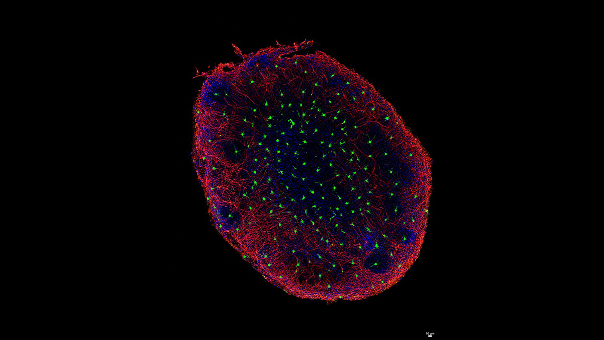
Figure: Human brain organoid containing microglia. Microglia are shown in green and neurons in red, illustrating the integration of brain immune cells within a dense neuronal network. Nuclei are shown in blue.
Mitochondrial Unfolded Protein Response Promotes Human Microglial Senescence
Proteostasis, the maintenance of a healthy, correctly folded protein network, is essential for cellular function. When mitochondrial proteins are damaged or misfolded, cells activate the mitochondrial unfolded protein response (UPRmt), a quality-control program that induces chaperones and proteases to help restore mitochondrial proteostasis. In simple model organisms, mild UPRmt activation has been linked to increased stress resistance and longer lifespan. A recent study led by María José Pérez and Michela Deleidi asked whether this response is equally protective in the human brain. The findings were published in Nature Neuroscience.
The team wondered how mitochondrial proteostasis is maintained in the aging human brain, how mitochondrial proteostasis is maintained in different human brain cell types and whether UPRmt activation has the same consequences across neurons and glia. Using human induced pluripotent stem cell-derived neurons, astrocytes and microglia, as well as neuronal-glial tricultures and microglia-containing brain organoids, they modeled mitochondrial proteotoxic stress in a human cellular context. Pharmacological inhibition of the mitochondrial protease LONP1 was used to induce mitochondrial protein misfolding and activate the UPRmt. This stress caused mitochondrial fragmentation and reduced mitochondrial membrane potential across cell types, but the downstream responses were markedly different.
The team found that mitochondrial stress profoundly altered human microglial metabolism, causing lipid remodeling, lipid-droplet accumulation and depletion of S-adenosylmethionine (SAM), a key metabolite involved in methylation and antioxidant defense. These changes were associated with DNA damage, inflammatory signaling and increased senescence. By contrast, neurons and astrocytes mounted more adaptive stress responses, whereas microglia showed impaired quality control and accumulated misfolded proteins.
The study shows that mitochondrial stress responses are highly cell-type specific in the human brain. Although the UPRmt can support proteostasis under some conditions, chronic activation in microglia can become maladaptive, promoting senescence and disrupting neuronal homeostasis. These results suggest that therapeutic strategies aimed at modulating mitochondrial stress responses will need to be carefully tuned by cell type, duration and disease context.
Statement of Significance by Drs. Pérez and Deleidi
Reports on the mitochondrial unfolded protein response, or UPRmt, have been contradictory, with protective effects in some settings and damaging effects in others. Our findings suggest that this depends, at least in part, on cellular context. Brain cells do not mount a uniform mitochondrial stress response: microglia responded earlier and more strongly than neurons, suggesting that they may act as key sensors of mitochondrial stress.
In microglia, however, sustained UPRmt activation was maladaptive, driving metabolic dysfunction, senescence and impaired communication with neurons and astrocytes. Microglia carrying a mitochondrial proteostasis defect were sufficient to induce senescence, reduce neuronal integrity and increase amyloid accumulation in brain assembloids. This signature overlapped with disease-associated microglial states found in aging and Alzheimer’s disease brain. These findings reveal how defects in mitochondrial quality control may contribute to neurodegeneration by altering the brain’s immune environment. They also highlight rare mitochondrial diseases as powerful models to uncover mechanisms relevant to common age-associated disorders, including Parkinson’s and Alzheimer’s disease. Overall, our study identifies microglial metabolism and mitochondrial proteostasis as potential therapeutic targets and underscores the need for cell-type-selective approaches rather than indiscriminate activation of mitochondrial stress pathways.
A Discussion with Drs. Pérez and Deleidi
MitoWorld: Can you give us an idea of where you might direct your future research to expand on the findings in this paper?
Authors:
One important direction will be to understand whether the mechanisms we identified in human microglia also operate in the context of specific neurodegenerative diseases. In this study, we found that chronic activation of the mitochondrial unfolded protein response can push microglia toward a senescent and inflammatory state. We now want to explore how this process interacts with disease-associated protein aggregates, such as α-synuclein in Parkinson’s disease or amyloid and tau pathology in Alzheimer’s disease.
We are also interested in defining whether the metabolic vulnerabilities we identified, particularly lipid remodeling and the SAM-polyamine axis, can be targeted to restore healthier microglial function. Ultimately, our goal is to understand whether correcting mitochondrial stress responses in microglia could help preserve neuronal homeostasis and slow disease progression.
MitoWorld: It is interesting that the mitochondria in different cell types had such different responses. Do you have any thoughts on why that might be? Is this just another case of how mitochondria are regulated by the nuclear genes?
Authors:
Yes, nuclear regulation is certainly part of the answer, but we think the explanation is broader. Mitochondria are deeply shaped by the identity, function and metabolic state of each cell type. Neurons, astrocytes and microglia have very different energetic demands, stress-response programs and roles within the brain. Therefore, the same mitochondrial stress can be interpreted very differently depending on the cellular context.
In our study, neurons and astrocytes appeared better able to activate adaptive compensatory pathways, whereas microglia showed a more maladaptive response, with impaired proteostasis, metabolic rewiring and senescence. This suggests that mitochondrial stress responses are not uniform across the brain. They are integrated with cell-type-specific transcriptional programs, metabolic wiring and immune functions.
MitoWorld: Each neurodegenerative disease features its own misfolded protein (e.g., Parkinson’s disease, a-synuclein; Huntington’s disease, huntingtin) and affects specific neurons. Could those be other examples of the type of cell-specific results of the UPR?
Authors:
Absolutely. One of the important lessons from our study is that proteostatic stress responses are highly cell-type specific. Different brain cells may have different thresholds for coping with misfolded proteins, and different diseases may expose vulnerabilities in distinct cellular populations.
In Parkinson’s disease, for example, dopaminergic neurons are particularly vulnerable, but microglia and astrocytes also shape how α-synuclein pathology spreads and how the tissue responds. Similarly, in Huntington’s disease or Alzheimer’s disease, the affected neurons and glial cells may engage different stress-response pathways depending on their metabolic state, proteostatic capacity and local environment. We think that understanding these cell-specific responses will be essential for designing therapies that do not simply activate or inhibit a pathway globally, but modulate it in the right cell type and at the right time.
MitoWorld: Do you have any speculation on the mechanism involved in the lipid remodeling?
Authors:
Our data suggest that lipid remodeling is not just a secondary consequence of mitochondrial stress, but part of the mechanism driving microglial dysfunction. When mitochondrial proteostasis is impaired, microglia appear to reorganize their lipid metabolism, leading to the accumulation of lipid droplets and changes in glycerophospholipid and glycerolipid pathways. One possibility is that lipid droplets initially form as a protective response to buffer damaged membranes, oxidative stress or excess fatty acids. However, when the stress is chronic, this adaptive response may become maladaptive. Lipid-droplet accumulation has been linked to impaired phagocytosis, increased inflammatory signaling and reduced cellular fitness in aging microglia. In our models, interfering with lipid-droplet formation reduced senescence markers, suggesting that lipid remodeling actively contributes to the senescent phenotype.
MitoWorld: As you note in the Discussion, your findings have significant implications for cancer treatments? Do you have plans to follow up on these possibilities?
Authors:
This is an important point. Some therapeutic strategies, including in oncology, aim to manipulate mitochondrial stress responses or proteostasis pathways. Our findings suggest that these approaches may have cell-type-specific effects that need to be carefully considered. In particular, chronic activation of mitochondrial stress responses in immune cells could potentially promote senescence or inflammatory dysfunction. Our main focus remains neurodegeneration, but we think the broader implication is that mitochondrial stress pathways should be studied in a tissue- and cell-type-specific manner. It will be important to understand when activation of the UPRmt is beneficial and when it becomes maladaptive. This principle could be relevant not only for brain diseases, but also for cancer and other age-associated conditions in which senescence, inflammation and mitochondrial dysfunction intersect.
MitoWorld: Can you tell us how you came to be interested in mitochondria?
Authors:
Our interest in mitochondria came from trying to understand why certain brain cells are especially vulnerable in neurodegenerative diseases. Mitochondria sit at the intersection of several processes that are central to these diseases: energy metabolism, inflammation, protein quality control, oxidative stress and cellular aging. For our group, mitochondria became particularly interesting because they provide a way to connect rare genetic disorders with broader mechanisms of neurodegeneration. Studying defects in mitochondrial proteostasis can reveal fundamental biological pathways that may also be relevant to common age-associated diseases. Over time, this led us to focus not only on neurons, but also on glial cells, especially microglia, and to ask how mitochondrial stress in these cells affects the entire brain environment.
Reference
Perez J MJ, Lam A, Weissleder C, Bertoli F, Raji H, Bosch M, Nemazanyy I, Kalb S, Kehili M, Hirschberg I, Brunetti D, Heckenbach I, Scheibye-Knudsen M, Deleidi M (2026) The mitochondrial unfolded protein response in human microglia disrupts neuronal–glial communication and promotes senescence. Nature Neuroscience https://doi.org/10.1038/s41593-026-02320-1.