The Great Mitochondrial Replacement
Cytotoxic T cells are vital for eliminating cancer cells and thus for restraining tumour growth. However, many types of cancer cell blunt this immune attack by issuing signals that somehow cripple the attacking cell. Like almost everything in immunity, this undoubtedly has multiple dimensions. However, one crucial aspect seems to be centred on mitochondria. By rendering the mitochondria of the cytotoxic cell incapable of energizing the process(es) that kill the target cell, cancer cells survive immune attack. This concept has received support from studies showing that T cells that invade the tumour micro-environment exhibit signs of mitochondrial energetic dysfunction. However, the cellular and molecular mechanisms behind this phenomenon have, until recently, remained obscure.
This phenomenon could have serious consequences. Cancer cells frequently harbour mtDNA mutations that impair their own capacity for oxidative ATP generation (OXPHOS). Surprisingly, mitochondria from T cells use ‘tunnelling nanotubes,’ to transfer into cancer cells [2-4] and promote tumour survival. Even if only a small fraction of tumour cells are ‘empowered’ in this manner, it should be sufficient to protect them from cytotoxic drugs that target cells dependent on non-mitochondrial pathways for their energy supply, DNA repair and cell division. Thus, a pool of chemoresistant cells would survive to reconstitute the tumour in an even more aggressive form that is refractory to future treatments. The phenomenon of intercellular transfer of mitochondria remains somewhat controversial. However, it has been independently documented by multiple reputable laboratories and must now be considered physiological. Nevertheless, it could still be an enhanced property of tumour cells or other cells with metabolic impairment.
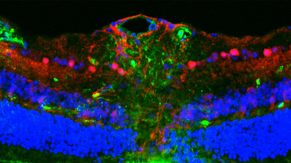
A recent paper from the lab of Yosuke Togashi in Japan shed light on one important part of this puzzle [1]. They report that mitochondrial transfer also occurs in the opposite direction, via tunnelling nanotubes and or/extracellular vesicles. OXPHOS-incompetent mitochondria from the tumour, bearing deleterious mutations in mtDNA, were detected in T cells that had invaded the tumour micro-environment. The origin of these mitochondria was confirmed by co-culture experiments with tumour cells expressing a mitochondrially targeted fluorescent reporter protein. Prolonged co-culture resulted in the almost complete replacement of endogenous T-cell mitochondria by those derived from the tumour cells. This replacement reflected the fact that tumour-derived mitochondria were resistant to mitophagy, whereas the original T-cell mitochondria were not.
T cells whose mitochondria had suffered this replacement showed OXPHOS impairment, increased dependence on non-mitochondrial ATP generation, downregulation of T-cell markers associated with cytotoxic functions, and signs of cellular senescence. Importantly, the acquisition of tumour- derived mitochondria impaired their activation and, hence, their ability to kill cancer cells, as shown by an in vivo model.
The work breaks new ground by showing that the interaction of immune cells with cancer cells can facilitate a bidirectional mitochondrial exchange that simultaneously favours cancer cell viability and immune evasion. However, this obviously raises many questions. The authors showed that the exchange can operate in at least three cancer-cell types (melanoma, breast and lung cancer), but does it work for all cancer types? Does the phenomenon depend on which surface markers are expressed by a given cancer cell, or by which antigen-receptors are expressed on the cytotoxic cell with which it interacts? The immune cell repertoire includes many different cell types, each with a role in anti-cancer immunity. Does mitochondrial exchange operate only in cytotoxic T cells, or is it seen in other immune cell-types, such as Treg and other CD4+ cells, dendritic cells, NK (natural killer) cells, phagocytes or B lymphocytes? If so, how is their function modified? Are those other immune cell functions secondarily deranged by impaired cytokine secretion from OXPHOS- deficient T cells? Does tumour mtDNA migrate to other cells in the body than immune cells, and if so, is their function also compromised?
The discovery of the mitochondrial fusion/fission cycle forced us to rethink the mitochondrial content of each cell as being akin to a single entity. The pioneering work of the Togashi lab and others now obliges us to revise another fundamental tenet of mitochondrial biology, recognizing that mitochondria and their DNA are not simply the chattels of single cells, but belong to populations of many cells and to tissues, organs and even the body as a whole.
References
- Ikeda H, et al. (2025) Immune evasion through mitochondrial transfer in the tumour microenvironment. Nature 638: 225–236. doi: 10.1038/s41586-024-08439-0.
- Tan AS, et al. (2015). Mitochondrial genome acquisition restores respiratory function and tumorigenic potential of cancer cells without mitochondrial DNA. Cell Metab. 21: 81-94. doi: 1016/j.cmet.2014.12.003.
- Saha T, et al. (2022) Intercellular nanotubes mediate mitochondrial trafficking between cancer and immune cells. Nat Nanotechnol. 17: 98–106. doi: 10.1038/s41565-021-01000-4.
- Zhang H, et al (2023) Systematic investigation of mitochondrial transfer between cancer cells and T cells at single-cell resolution. Cancer Cell 41: 1788–1802.e10. doi: 10.1016/j.ccell.2023.09.003.