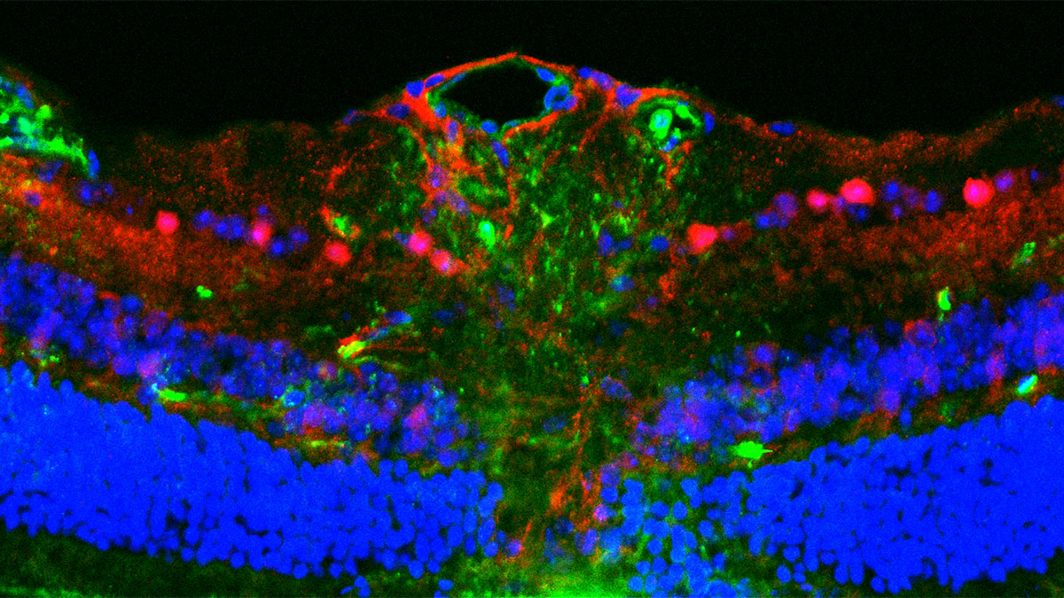
The Mitochondrial Intermembrane Space
Mitochondria consist of an outer membrane, a highly folded inner membrane, an inner matrix, and an intermembrane space (IMS). In an excellent recent review, Fara van der Schans, Kostas Tokatlidis, and Daniela G. Vitali of the University of Glasgow describe the mitochondrial IMS, its proteome, and functions. The review was published in the journal Protein Science.
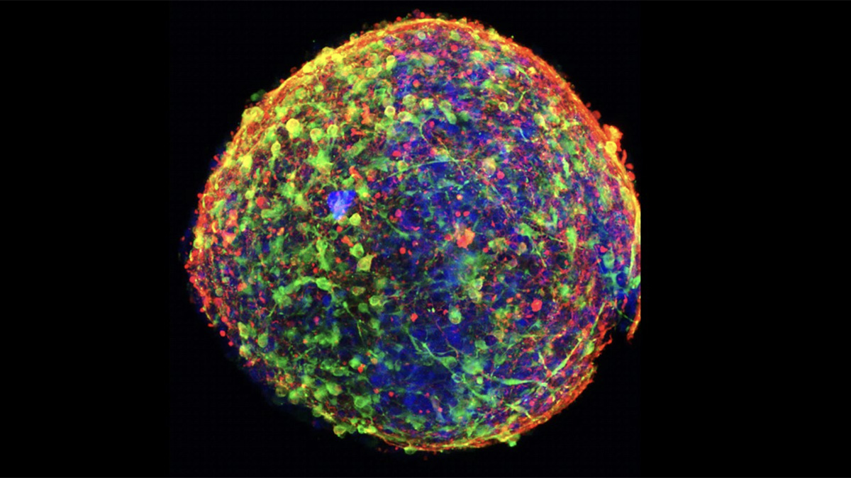
The IMS lies between the outer and inner membranes. This location makes it a key transit area for the movement of proteins and other material and makes it a signaling hub for the mitochondria. In fact, nuclear genes encode all of the proteins in the IMS (about 130 proteins). Those proteins are imported into the IMS by various pathways, including the mitochondrial intermembrane space assembly import (MIA) pathway, stop-transfer pathway, and other non-canonical mechanisms. They are important in various cellular pathways (e.g., redox regulation, calcium signaling, apoptosis, and hypoxia response). In addition, most mitochondria-encoded proteins pass through the IMS on the way to their final destinations.
About a third of IMS proteins are imported via the MIA pathway. A number of specific topologies are involved in the transport, and those proteins are then folded in the IMS to become mature proteins. The coordination and targeting of nuclear and mitochondrial proteins are complex operations. There is a high risk of mutations, misfolding, and damage from reactive oxygen species that are produced in mitochondria. A mitochondrial quality control system helps to protect against these risks. Together, these mechanisms maintain cellular homeostasis and mitochondrial function.
This is an excellent overview of the IMS. It carefully examines the path of proteins into and through the mitochondria as they are imported, processed, reach their site of function, and eventually are exported back out of the mitochondria to the proteasome for degradation.
A Statement of Significance from Dr. Tokatlidis:
Understanding how proteins are targeted, sorted, and monitored within the mitochondrial intermembrane space (IMS) is crucial because this compartment acts as a central checkpoint for mitochondrial proteostasis. Although small in volume, the IMS oversees the maturation and quality control of nearly all mitochondrial proteins as they traverse the organelle, ensuring that only correctly folded and functional proteins proceed to their destinations while harmful intermediates are removed. This surveillance is vital for maintaining mitochondrial integrity, as disruptions in IMS protein handling can compromise respiration, redox balance, apoptosis, calcium signaling, and cellular stress responses. By synthesizing recent insights into IMS protein import and quality-control pathways, this review clarifies how this compartment integrates protein sorting with broader mitochondrial homeostasis. Appreciating the IMS as an active regulatory node highlights its fundamental role in safeguarding mitochondrial function and underscores why its dysregulation contributes to diverse human diseases, from neurodegeneration to metabolic disorders.
A Conversation with Dr. Tokatlidis:
MitoWorld: This is an excellent review of the structure and functions of the ISM. Can you give us an idea of how your research relates to findings in this review and where you are going next?
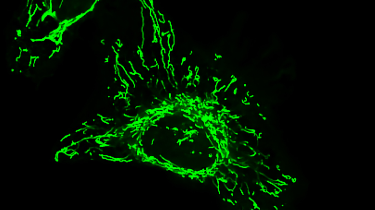
Dr. Tokatlidis We have been studying for a number of years now how proteins get to the IMS initially focusing on the stop transfer pathway and then on the discovery and characterization of the MIA pathway. Our work on the MIA import and assembly system started with a surprising finding for the small Tim chaperone proteins. These small proteins (about 10 kDa) form essential chaperones assemblies in the IMS that are critically needed to assist membrane proteins negotiate their passage across the aqueous IMS. In the absence of these dedicated chaperones membrane proteins like the metabolite transporters (including the ADP/ATP transporter) and even proteins of the outer membrane on their way to be inserted would aggregate and become nonfunctional, with detrimental effects for mitochondria. We wondered how these small TIM chaperones themselves get imported, as they do not have any sort of mitochondrial targeting sequence. We found to our surprise that a critical step in the import, folding and retention of these proteins in the IMS is the formation of internal disulfide bridges once they are imported into the IMS, which indicated that this compartment has the capacity for generating disulfide bonds in proteins (the process of oxidative folding). This was quite a significant departure from the prevailing dogma that disulfide bonds in eukaryotic cell can only be generated in the endoplasmic reticulum and was not widely accepted for a couple of years. The work of several groups, including ours, then showed that the protein Mia40 is the key player to catalyse the process of oxidative folding in the IMS. We further went on to define the structure of Mia40, how it interacts in transient manner with its import substrates, the unusual targeting signal that underpins this pathway and is very different from the conventional mitochondrial signals and the mechanistic features and structural and thermodynamic basis of the interactions that guide this IMS import pathway. The MIA pathway is unique among all import pathways into mitochondria as it is the only one that chemically modifies the imported protein by introducing covalent bonds (disulfide bridges). This is pathway is also linked to redox control in mitochondria and the more general redox homeostasis cues in the cell, so it is critical to understand the links between this aspect of mitochondria biogenesis and redox balance. This is where our research focuses next. We want to understand how redox balance is maintained in the IMS and how this is connected to cellular redox signalling, particularly looking for non-conventional import pathways of antioxidant proteins that make their way to the IMS and how these processes work in stress conditions. Recent work in our lab with pancreatic cancer cells shows that such pathways may hold the key to understanding vulnerabilities in come of the most therapy resistant cells and we want to tease out these links. We are also interested in establishing the crosstalk between these redox-regulated mitochondria biogenesis processes and mitochondria dynamics and contacts with other organelles, which are at the heart of mitochondria function and mitochondrial damage.
MitoWorld: We are always intrigued by how the nuclear and mitochondrial genes interact to share the genetic control of the cell. The IMS seems like a location where much of this cooperation is manifested. Do you have any thoughts on this?
Dr. Tokatlidis Yes, it is true that the IMS is like a buffer zone between the cytosol and the inner most mitochondrial compartment, the matrix. Because of the presence of porins in the OM, small molecules up to about 5 kDa can freely diffuse in and out of the IMS. This allows several signalling molecules to contribute to the communication between the mitochondria core and the cytosol. Additionally, the IMS is the compartment that discharges to the rest of the cell several of the apoptosis factors upon selective permeabilization of the OM in conditions of programmed cell death. A very fine regulation of the IMS both structurally and functionally in response to changing metabolic and stress conditions is therefore fundamental to control such processes and allow the function of mitochondria as a signalling organelle. All of the IMS proteins are nuclear-encoded, but many of them act as assembly factors of the OXPHOS complexes in the IM, which contain subunits encoded by the mtDNA. It is therefore critical that the coordination of these interactions in the IMS works well to allow assembly and stability of OXPHOS and eventually maintenance of mitochondrial fitness.
MitoWorld: Can you speculate on the signaling mechanisms that facilitate that joint control of cellular activities?
Dr. Tokatlidis All current research points to the direction that the IMS acts as a signaling nexus rather than a passive compartment and is able to coordinate and integrate cellular activities. The IMS sits at a fascinating intersection: chemically oxidizing, topologically distinct and structurally constrained. That combination makes it uniquely suited for rapid and reversible signaling. Some of the several mechanistic “modules” that plausibly operate together to allow the IMS to exert joint control over cellular activities are: (1) Import flux as a proxy for cellular metabolic state, (2) Mia40 oxidation cycles encoding redox information, (3) IMS glutathione pool (more oxidized than matrix) acting as a tunable buffer, (4) H₂O₂ microdomains near respiratory complexes acting as localized second messengers (5) protein conformational switching as a signaling currency, (6) IMS proteins may act as electrochemical translators, converting changes in Δψ or proton leak into structural or redox signals that regulate OM channels (VDAC, BAX/BAK priming, TOM complex dynamics). (7) the IMS may regulate OM mechanotransduction, where changes in cristae architecture alter OM tension, influencing cytosolic signaling platforms, such as cytosolic Ca²⁺ signaling, metabolic fluxes (ATP/ADP exchange), apoptotic priming, innate immune signaling (e.g., MAVS activation indirectly via OMM tension and ROS), (8) the IMS redox and structural states may be decoded by cytosolic kinases or ubiquitin ligases that sense OM conformational changes, forming a mechano-redox signaling axis, and finally (9) cells may use “IMS leakage pulses”—brief, reversible permeability events—to communicate mitochondrial stress without committing to apoptosis.
MitoWorld: The quality control of proteins as they are imported and processed is quite complex to maintain a delicate balance involving chaperones, modifiers, and degradation in the proteosome. Much of this takes place in the IMS. Can you elaborate on that balance?
Dr. Tokatlidis Mitochondrial protein quality control hinges on a finely tuned equilibrium in which the IMS acts as a critical decision point, coordinating chaperone buffering, oxidative folding, and degradation. As precursors enter the IMS, small TIM chaperones and the Mia40–Erv1 oxidative folding system manage their stability, ensuring hydrophobic or cysteine‑rich substrates remain import‑competent while avoiding over‑oxidation that traps misfolded intermediates. The IMS redox environment, more oxidizing than the cytosol or matrix, sensitively dictates whether proteins fold productively, undergo corrective modification, or are flagged for removal. Stalled or misfolded IMS intermediates can be retro‑translocated for cytosolic proteasomal degradation or passed inward to matrix chaperones and proteases, but excessive degradation risks depleting essential IMS components and disrupting respiratory chain assembly. The overall balance emerges from the interplay of IMS redox poise, chaperone capacity, import flux, and protease thresholds, making the IMS a central hub for maintaining mitochondrial proteostasis under fluctuating metabolic and stress conditions.
MitoWorld: We are always interested in what attracted researchers to mitochondria. How did you become interested in them in the first place?
Dr. Tokatlidis I was trained as a chemical engineer during my undergraduate studies and became fascinated by how interdisciplinary research can be used to tackle major scientific problems. This interest led me to pursue a PhD in biochemistry, where I worked on protein folding and the functions of molecular chaperones. In the era before AlphaFold, protein folding was considered essentially intractable, and even now, despite enormous advances, it remains a highly complex challenge for cells.
My growing interest in the principles of protein folding and assembly naturally drew me toward mitochondria, given their central importance in cellular life. I was fortunate to join the laboratory of Jeff Schatz at the Biozentrum in Basel. Jeff was a towering figure in mitochondrial biology—the co‑discoverer of mitochondrial DNA in the 1960s and a pioneer in elucidating the mitochondrial protein import machinery. Working with him had a profound influence on me and shaped the entire trajectory of my scientific career.
Following the discoveries I made early on, I have remained committed to mitochondrial research for nearly 30 years. Throughout my career—in Manchester, then in Crete, and now in Glasgow—I have been lucky to work with exceptionally engaged and driven colleagues from around the world. Together, we have explored fundamental pathways that not only deepen our understanding of mitochondria but also provide a foundation for translational applications in health and disease.
I believe mitochondrial biology is central to understanding many common human pathologies, including cancer, neurodegeneration, and cardiovascular disease, as well as rare mitochondrial disorders and the biology of ageing. One of the most rewarding aspects of working in this field is the constant sense of discovery: mitochondria continually surprise us with their involvement in nearly every aspect of cellular homeostasis.
Finally, collaborating with talented and motivated young researchers has made this journey even more exciting, and it remains one of the most fulfilling parts of my work.
Reference
van der Schans F, Tokatlidis K, Vitali DG (2026) In and out of the mitochondrial intermembrane space. Protein Science 35(3): e70493.