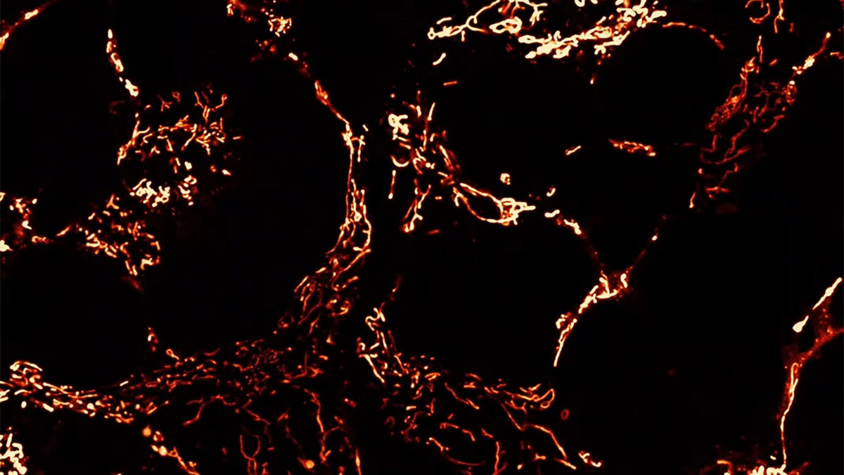
Mitochondria labeled with MitoTracker orange in human embryonic kidney cell culture illustrate the dynamic architecture of the mitochondrial network and its continuous remodeling. Image credit: Oleg Kovtun, PhD and Andrea Marshall, PhD.wo
MICOS Complex May Contribute to Mitochondrial Decline During Kidney Aging
Aging kidneys exhibit mitochondrial structural defects, oxidative stress, fibrosis, and altered metabolism. A new study published in Aging Cell suggests that alterations in the mitochondrial contact site and cristae organizing system (MICOS) may contribute to these defects.
Kidney function commonly declines with age, and aging is a major risk factor for acute and chronic kidney disease. Because many kidney cell types, especially tubular epithelial cells, have high energetic demands and rely heavily on mitochondrial function, the research team asked whether age-related changes in mitochondrial architecture contribute to this vulnerability.
Using transmission electron microscopy (TEM) and serial block-face scanning electron microscopy (SBF-SEM), the investigators examined mitochondria from the kidneys of young versus old mice. In the aged cohort, they found fragmented mitochondrial morphology, and, notably, the folds of the inner mitochondrial membrane that promote energy production, known as cristae, were disorganized, shorter, and smaller by volume. At the molecular level, aging was correlated with decreased expression of MICOS components and of OPA1, another key regulator of inner mitochondrial membrane structure.
To test whether MICOS disruption could affect mitochondrial function, the authors perturbed MICOS components in cellular models. Loss of MICOS proteins altered mitochondrial calcium handling, increased reactive oxygen species, and impaired aspects of mitochondrial respiration. Human genetic and biobank analyses further linked CHCHD6 and OPA1 with kidney and genitourinary disease phenotypes, although these findings remain associative.
Together, the study supports a model in which age-related decline of MICOS and related cristae regulators contributes to deterioration of mitochondrial inner membrane architecture. These changes are associated with oxidative stress, altered calcium regulation, metabolic remodeling, fibrosis, and increased vulnerability to kidney dysfunction during aging.
A Statement of Significance from Dr. Hinton, Jr.:
Aging is the greatest risk factor for chronic kidney disease, yet the mechanisms linking aging to renal dysfunction remain poorly understood. In this study, we demonstrate that aging is associated with profound remodeling of mitochondrial ultrastructure in the kidney, characterized by disrupted cristae architecture, altered mitochondrial morphology, increased oxidative stress, and widespread metabolic dysregulation. Using advanced three-dimensional electron microscopy, human kidney samples, large-scale biobank analyses, and mechanistic studies targeting the mitochondrial contact site and cristae organizing system (MICOS) complex, we identify age-dependent loss of MICOS components as a conserved feature of kidney aging. Furthermore, disruption of MICOS proteins directly impairs mitochondrial function, calcium handling, and redox homeostasis, recapitulating key features of the aged kidney phenotype. These findings establish MICOS complex dysfunction as a central regulator of mitochondrial structural integrity during renal aging and suggest that preserving cristae architecture may represent a therapeutic strategy to mitigate age-related kidney disease and preserve renal healthspan.
A conversation with the authors:
MitoWorld: What initially led your group to investigate the MICOS complex during kidney aging?
Hinton et. al.: This study grew from a broader question that has guided our work for many years: Does mitochondrial architecture actively influence aging, or is it simply a consequence of aging? For decades, mitochondrial dysfunction has been widely recognized as a hallmark of aging, but most studies focus on metabolism, ATP production, and oxidative stress. The primary focus was on the functional aspect, with the structural aspect given a back seat. We wanted to determine whether mitochondrial structural organization also contributes to tissue decline. This study draws the field’s attention to the role of structural organization in cellular aging.
Our laboratories had previously observed age-associated declines in MICOS proteins in skeletal and cardiac muscle, along with disrupted cristae organization, altered mitochondrial morphology, oxidative stress, and metabolic dysfunction. These recurrent observations from our lab and elsewhere suggested that MICOS decline might represent a broader biological phenomenon rather than a tissue-specific event. The kidney was compelling because it is highly energetic and densely packed with mitochondria. Even subtle changes in mitochondrial architecture could have major consequences for kidney function. Also, independent human genetic observations from our lab and Dr. Katti’s lab provided additional evidence supporting the rationale for investigating MICOS biology in renal aging.
MitoWorld: What was the major discovery of the paper?
Hinton et. al.: The major discovery was that kidney aging is accompanied by a breakdown of mitochondrial architecture, closely linked to the loss of the MICOS complex. We observed coordinated declines in MIC60, CHCHD3, CHCHD6, MIC10, MIC13, and OPA1—proteins that help maintain cristae and inner membrane organization. The key distinction from previous reports in our study is that this architectural maintenance system appears to deteriorate systematically during kidney aging.
These changes were associated with cristae disorganization, altered mitochondrial networks, oxidative stress, calcium dysregulation, fibrosis, and metabolic remodeling. Mechanistically, we propose that MICOS loss destabilizes cristae, reducing electron transport efficiency and promoting reactive oxygen species production. This may create a feed-forward cycle of mitochondrial damage. The key conceptual leap and contribution is that age-related loss of MICOS-dependent mitochondrial architecture may be a central organizing feature that links cristae disruption, mitochondrial remodeling, oxidative stress, metabolic dysfunction, and altered organelle communication in the aging kidney.
We also observed age-associated changes in mitochondria–endoplasmic reticulum contacts; the novelty here is not simply cristae disruption, but the suggestion that age-related architectural decline may influence communication between organelles.
MitoWorld: How do alterations in cristae architecture contribute to oxidative stress and mitochondrial dysfunction during aging?
Hinton et. al.: Some of the most transformative discoveries in biology emerged from the realization that structure can be a mechanism rather than merely a consequence of function. The double-helical architecture of DNA explained heredity, the quaternary structure of hemoglobin revealed cooperative oxygen transport, the organization of the sarcomere uncovered the basis of muscle contraction, and the intricate architecture of the ribosome illuminated the process of protein synthesis. In each case, biological form provided the missing causal link between molecular organization and physiological outcome.
Emerging research suggests that mitochondrial cristae follow this same pattern. Rather than treating age-associated changes in cristae morphology as a passive hallmark of mitochondrial decline, we seek to address whether alterations in cristae architecture can themselves drive functional deterioration, for example, in altered electron transport, oxidative stress, and impaired cellular and organ physiology.
Cristae organize the respiratory machinery required for oxidative phosphorylation. In aged kidneys, we observed reduced cristae volume, surface area, and integrity, accompanied by decreased MICOS expression. Disruption of cristae architecture likely impairs electron transport efficiency, increases electron leakage and reactive oxygen species production, and promotes oxidative damage. Because cristae also regulate respiratory supercomplex assembly, calcium homeostasis, and organelle communication, their deterioration may contribute broadly to mitochondrial and cellular dysfunction during aging.
MitoWorld: What advantages did 3D electron microscopy provide compared with traditional two-dimensional approaches?
Hinton et. al.: At its core, this question asks what level of observation is necessary to reveal the biological organization of aging mitochondria. A two-dimensional image can tell us what an individual mitochondrion looks like, but aging is unlikely to be an isolated organelle phenomenon. We wanted to understand how mitochondrial networks are organized throughout a tissue and how those relationships change with age. Three-dimensional electron microscopy allowed us to observe mitochondria in their native architectural context, revealing that aging may involve a loss of network organization and organelle connectivity, rather than simply damage to individual mitochondria. 3D EM allows aging to be studied as a network-level architectural process rather than as isolated mitochondrial snapshots.
Using serial block-face scanning electron microscopy, we reconstructed mitochondrial networks throughout kidney tissue. This allowed us to quantify volume, surface area, connectivity, branching, network organization, and spatial relationships with nearby organelles. A major advantage was that 3D imaging revealed network remodeling and connectivity loss that were not apparent from isolated 2D views. It also allowed us to examine how mitochondria are positioned relative to the endoplasmic reticulum, supporting our interest in the mitochondrial connectome.
MitoWorld: Do you believe MICOS proteins represent viable therapeutic targets for age-related kidney dysfunction?
Hinton et. al.: One way to think about this question is not simply whether MICOS proteins can be targeted therapeutically, but whether preserving mitochondrial architecture can preserve tissue function during aging. MICOS proteins sit at the intersection of structure and function, organizing cristae architecture, inner membrane integrity, respiratory supercomplex assembly, calcium handling, and interactions with proteins such as OPA1. This makes them particularly attractive because they may act upstream of many aging-associated processes rather than regulating a single pathway.
In our study, MICOS decline was associated with cristae deterioration, oxidative stress, metabolic remodeling, fibrosis, and kidney dysfunction. However, the critical unanswered question is whether restoring MICOS function can reverse existing pathology or merely slow its progression. Ultimately, the therapeutic promise of MICOS depends on whether mitochondrial architecture is a driver of aging or simply a marker of it.
MitoWorld: What are the most important unanswered questions regarding MICOS biology in aging tissues?
Hinton et. al.: Perhaps the most important unanswered question is not why a particular MICOS protein declines with age, but whether biological architecture itself represents a fundamental layer of aging biology. We have spent decades cataloging age-related changes in genes, proteins, and metabolites, yet cells are also highly organized spatial systems.
The emerging challenge is to determine whether aging reflects a gradual loss of this organization and whether restoring architectural integrity can restore function. Addressing that question will require us to move beyond studying individual organelles and toward understanding the cellular connectome—the network of interactions that links mitochondria with the ER, lysosomes, lipid droplets, and other compartments. In that sense, the future of the field may be less about mitochondria themselves and more about how cellular architecture is maintained across the lifespan.
Looking ahead, we believe the most exciting aspect of this work is not what it answers, but what it leaves unanswered. In our opinion, the following questions will be particularly important in the years ahead, as they address the fundamental relationship between mitochondrial architecture, cellular organization, and aging.
- What drives the age-dependent decline of the MICOS complex?
- Can restoration of MICOS reverse age-associated dysfunction?
- How does MICOS shape communication between mitochondria and the rest of the cell?
MitoWorld: What are the major limitations of the study?
Hinton et. al.: Every study ultimately comes down to the difference between what we can observe and what we can conclude. Scientific progress often occurs in two stages: first, identifying a pattern, and then proving that the pattern is causal. This study provides strong evidence that MICOS decline and architectural remodeling accompany kidney aging, but the ultimate test will be determining whether restoring that architecture can meaningfully alter the aging process itself.
Reference: