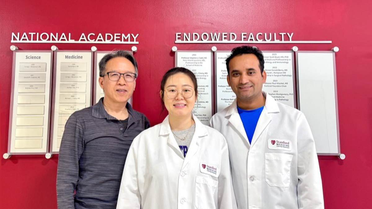
Photo of authors Bingwei Lu, Wen Li & Suman Rimal (left to right)
Tau Regulates Mitochondrial Reverse Electron Transport
For some time, tau has been an enigmatic protein. Aggregations of tau form the tangles reported by Alois Alzheimer as one of the features of Alzheimer’s disease. Tau is thought to stabilize microtubules and participate in other functions in brain neurons. However, pathological forms of tau (e.g., hyperphosphorylated tau, aggregations of tau) are associated with tauopathies, such as Alzheimer’s disease, frontotemporal dementia, progressive supranuclear palsy, and Pick’s disease. Yet, the link between tau and those diseases is unclear.
The team led by Dr. Lu sought to better understand that link. The results of their study were recently published in the journal Neuron.
The team used three experimental systems: flies, mice, and human iPSCs carrying a mutation that causes tau hyperphosphorylation and induced them to form neurons. By manipulating these cells, they found that reverse electron transport (RET) was enhanced. The result is that the cells produce more reactive oxygen species and have lower NAD+/NADH ratios. The researchers found that tau enters the mitochondria and interacts with complex 1 subunit NDUFS3. These actions cause increased RET and tau hyperphosphorylation.
The key finding of the study was to show that tau regulates RET, and that interfering with this this normal function can cause disease. It also found that inhibiting RET reduces tau levels and the risk of tau toxicity. Thus, regulating RET might be a promising therapeutic target.
A Statement of Significance by Dr. Lu
A major hurdle to understanding the core pathogenic mechanisms of tau and developing effective therapies is the uncertainty of the normal physiological function of tau that becomes altered in disease. This study provides across species evidence that tau plays a physiological role in facilitating mitochondrial reverse electron transport (RET), presumably as a stress adaptation mechanism. However, persistent RET activation by tau mutations, chronic stress, or aging becomes pathological, driven by a self-perpetuating vicious cycle in which phosphorylated tau (p-tau) promotes RET, and RET further promotes tau phosphorylation through tau kinases. Pharmacological inhibition of RET with a brain penetrant small molecule breaks the vicious cycle and offers therapeutic benefits in animal models and human iPSC derived neuronal models, nominating RET inhibition as a viable therapeutic approach for a broad spectrum of brain diseases that involve tau.
A Conversation with Dr. Lu
MitoWorld: Can you give us an idea of the direction you might take to advance the research contained in this paper?
Dr. Lu: This study opens many interesting research directions: 1) What is the normal physiological function of reverse electron transport (RET)? We know it is activated by stresses such as higher ambient temperature. But we know little about its physiological role in stress adaptation and response. 2) Other than tau, what other cellular proteins can modulate RET? 3) At the structural level, how does tau binding to mitochondrial complex I promotes RET, and how can this be inhibited by small molecule drug candidates? 4) How are the reverse and forward electron transport processes coordinated and differentially regulated? These are the research directions we are actively pursuing.
MitoWorld: Your work shows interaction with several complex systems (e.g., sirtuins, NAD+, ROS, post-translational modifications, protein aggregations). The inability to unravel all of these has thwarted research into Alzheimer’s disease for example. Can you speculate how these might fit together?
Dr. Lu: It is possible that the seemingly disparate, complex systems mentioned above may not be so isolated from each other after all. The fact that RET inhibition ameliorates disease phenotypes and modifies all these systems suggests that RET activation may sit at the top of the cascade and drives these different aspects of disease phenotypes. For example, RET generated ROS can activate tau kinases to promote tau hyperphosphorylation and aggregation. RET induced NAD+/NADH ratio change can alter cellular metabolism and impact the activities of signaling molecules such as the Sirtuins, which use NAD+ as co-factors. Previous efforts have targeted the downstream effector of RET individually, explaining their limited clinical success in Alzheimer’s disease and other neurodegenerative diseases.
MitoWorld: You mention that regulating RET might have clinical implications. Can you speculate on how that might be done, and do you have any plans to pursue such studies?
Dr. Lu: Our study indicates that RET acts early in the disease process and that RET and p-tau forms a self-perpetuating pathological loop that drives tauopathies. Our results also show that CPT, a small molecule that inhibits p-tau binding to mitochondrial complex I, breaks the RET/p-tau pathological loop and offers therapeutic benefits in multiple animal models and human iPSC-derived neuronal models of tauopathies. CPT belongs to Cerepeut, Inc, which is currently conducting IND-enabling studies on the compound.
MitoWorld: Can you tell us how you came to be interested in mitochondria and their roles in physiology and disease?
Dr. Lu: I first became interested in mitochondria when I was an undergraduate student at Fudan University where I did my thesis research on plant mitochondria. I was fascinated by mitochondria for their origin: The evolutionary origin of mitochondria is explained by the endosymbiotic theory, which suggests that mitochondria were once free-living prokaryotes that entered a symbiotic relationship with a host cell some billion years ago. This transformation from an independent organism to a specialized organelle is perhaps one of the most significant events in the history of life, because it enabled the development of complex multicellular organisms. Mitochondria are also fascinating because of their versatile roles in biology: They are best known as the power plant of cells due to their fundamental roles in making ATP, the currency of energy. But they do more than just making energy. It is becoming increasing clear that they are critical signaling organelles that control the proliferation, differentiation, and survival of the host cells. As a result, dysfunction of this organelle features prominently in diverse human diseases. Mitochondria are truly iconic organelles in biology and medicine.
My scientific career is intimately intertwined with mitochondria. I did my PhD at Cornall University, where I worked with Dr. Maureen Hanson on mitochondrial biology, specifically, RNA editing in plant mitochondria. For my postdoctoral training I wanted to do something different. I joined the Jan lab at UCSF, where I changed my research direction to neuroscience, studying the behavior of neural stem cells in fruit flies. After my postdoc, I established my independent lab first at the Rockfeller University in New York city and later at Stanford University, where in addition to continuing the work initiated during my postdoc, I started new research directions, working on the mechanism of brain diseases, from neurodegenerative diseases to brain tumor. It is the disease related research that led me to bump into mitochondria again. For example, our work on PINK1 and Parkin, two genes linked to familial Parkinson’s disease, and our work on the Notch signaling pathway in cancer biology have all implicated the fundamental involvement of mitochondria.