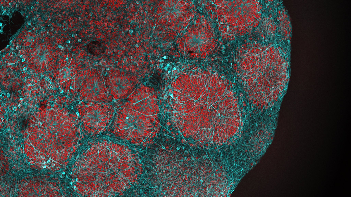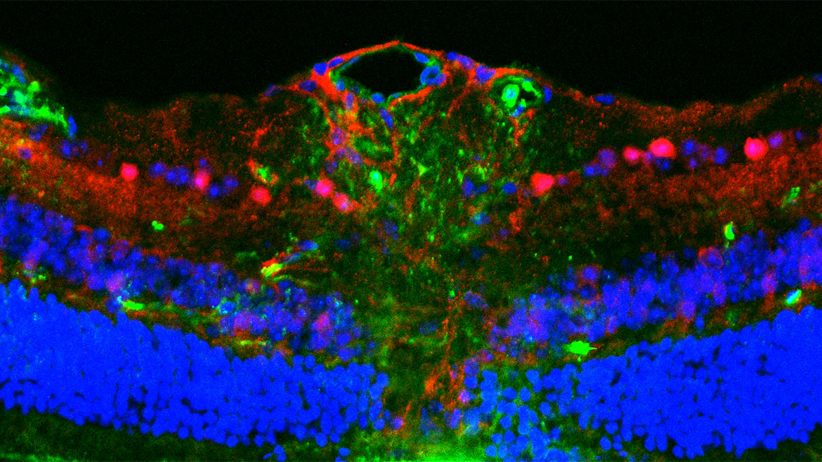
Immunofluorescence image of mouse optic nerve head with mitochondria (green), phospho-PGC-1⍺ (red) and nucleus (blue)
Metabolic Dysfunction in Glaucoma
A multi-institute research team, led by Arupratan Das, sought to find possible drugs to treat glaucoma. Using a high throughput mitochondrial screen in retinal ganglion cells (RGCs), they identified the 5-HT1A antagonist WAY-100635 (WAY) as an intriguing candidate. The paper was published in a recent issue of the journal Communications Medicine.
Early metabolic dysfunctions in RGCs have been implicated in glaucoma. Moreover, mitochondrial abnormalities cause degeneration of RGCs and have been implicated in mitochondrial optic neuropathies (MON), such as Leber hereditary optic neuropathy (LHON) and dominant optic atrophy (DOA).
Unfortunately, there is no approved therapy for preserving vision in this disorder. The Das research team set out to solve this problem. They used a live-cell mitochondrial screen in human embryonic stem cell-derived retinal ganglion cells. They identified the 5-HT1A antagonist WAY-100635 (WAY) as a candidate. WAY restored mitochondrial fitness, inhibited excitotoxicity, and enhanced aerobic glycolysis. It also preserved visual acuity and stopped glaucoma progression of the disease in mouse models of glaucoma mice. Importantly, WAY has already been approved for other indications, and not surprisingly, it showed no toxicity in the models used in this study.
The findings of this study are significant. No approved therapy is currently available for treating glaucoma. Here, the authors identified WAY as an intriguing possible treatment for this debilitating disorder. The results will give new hope to patients with glaucoma and other mitochondrial optic disorders.
A Statement of Significance from Dr. Das:
Glaucoma causes irreversible blindness by damaging retinal ganglion cells (RGCs), the neurons that carry visual information from the eye to the brain. Today’s treatments mainly lower eye pressure, yet many patients still lose vision because no approved therapy directly protects these neurons or preserves the retina-to-brain visual circuit. In this study, we used a live-cell screen in human stem cell–derived RGCs to identify a small molecule that strengthens cell’s energy producing unit mitochondria and protects these neurons. We show that it boosts key survival signaling, restores mitochondrial function, and preserves visual pathway activity in animal models. A major translational advantage is that this compound has already been used in human brain-imaging clinical studies, providing an existing safety foundation that could accelerate development of a first-in-class neuroprotective add-on therapy for glaucoma and potentially other optic nerve diseases.
A Conversation with Dr. Das:
MitoWorld: What are your plans to continue this line of research?
Dr. Das: This publication is the starting point for a much broader research program. Next, we want to determine which retinal ganglion cell (RGC) subtypes are protected; because different RGC classes support distinct visual functions (light/dark sensitivity, motion detection, and image-forming vs. non–image-forming vision). In parallel, we will deepen the molecular mechanism in human stem cell-derived RGCs by mapping the full downstream signaling network triggered by 5-HT1A antagonism, how cyclic adenosine monophosphate (cAMP) dynamics, PGC-1α–linked mitochondrial biogenesis, and mitochondria independent signaling converge to prevent degeneration. We will also define the metabolic reprogramming in the native retina, asking how the treatment reshapes energy use across compartments (RGC soma vs. long axons in the optic nerve), and whether this involves coupling mitochondria to aerobic glycolysis to sustain axonal structure and transport. Finally, we will test whether protection is purely RGC-autonomous or also involves neighboring retinal neurons, glia, and immune cells, and we will rigorously distinguish axon preservation from true axon regrowth with longitudinal tracing and optic-nerve profiling.
MitoWorld: Can you expand on the mechanisms for how WAY seems to protect the RGCs?
Dr. Das: In our study, we show that WAY acts by antagonizing the 5-HT1A G-protein–coupled receptor (GPCR), which reversibly elevates cyclic adenosine monophosphate (cAMP) in retinal ganglion cells. cAMP is a central second messenger that coordinates multiple neuroprotective programs, and in our system, it restores mitochondrial health in part by transiently activating PGC-1α–dependent mitochondrial biogenesis. Improving mitochondrial fitness showed two key consequences: it reduced the metabolic “workload” per mitochondrion (lowering stress while sustaining energy supply), and it supported a protective metabolic state that couples mitochondria with aerobic glycolysis. That glycolytic program is not just an ATP backup; it supplies building blocks needed for protein and lipid synthesis that help RGCs survive and maintain long-distance axonal structure and function.
MitoWorld: Your study was predicated on finding possible therapies for glaucoma. Do you have plans to take WAY into clinical trials?
Dr. Das: Yes, we are actively planning an FDA-enabling path and are serious about advancing WAY toward a first-in-human trial. Our next goal is to generate a complete Good Laboratory Practice (GLP) preclinical toxicology and pharmacokinetic data package using GLP-manufactured WAY, including dose-ranging, safety margins, and exposure–response relationships in a higher model (for example, canine), to support an Investigational New Drug (IND) application and a Phase I clinical trial. In parallel, we are optimizing drug formulation and delivery to maximize bioavailability and real-world usability, prioritizing an oral regimen and a long-acting depot option (for example, poly(lactic-co-glycolic acid) (PLGA) slow-release packaging for intramuscular administration) that could be practical across different patient age groups. We are also actively seeking funding to execute these critical translational studies because we believe this program represents a real opportunity to deliver a neuroprotective therapy for glaucoma, and potentially other optic neuropathies, where current treatment options remain limited to pressure management.
MitoWorld: You note that these findings might benefit studies of other neurodegenerative diseases. Can you elaborate on how that might work?
Dr. Das: The reason we believe these findings can extend beyond glaucoma is that WAY targets a core, convergent stress pathway that many neurodegenerative conditions share -loss of metabolic resilience, and mitochondrial stress. We are already testing this directly in traumatic brain injury (TBI) models and are seeing strong, reproducible early signals: TBI triggers progressive degeneration of retinal ganglion cells and robust immune activation in injured brain regions, whereas following injury WAY treatment preserves retinal ganglion cell survival, markedly reduces neuroinflammatory responses in the lesion, and is associated with improved visual function (including visual acuity) and reduced anxiety-like behavior. We are currently completing the remaining validation experiments and preparing this dataset for submission, with the broader goal of defining where this mechanism provides the greater therapeutic leverage across optic neuropathies and related brain-injury conditions.
MitoWorld: You mention that, in some glaucoma models, intraocular pressure disrupts mitochondrial function. Do you have any idea of how that happens?
Dr. Das: Yes. In glaucoma, high eye pressure puts the greatest strain on the point where retinal ganglion cell axons leave the eye, the optic nerve head. These axons are still unmyelinated there and they make a sharp turn as they enter the nerve, which makes this region mechanically and energetically demanding. Under pressure stress, axonal transport slows down, so mitochondria and other cargo can pile up in the optic nerve head. Because unmyelinated axons already rely heavily on mitochondria for energy, this added crowding and stress can increase harmful mitochondrial byproducts and damage the mitochondria, making the retinal ganglion cells more vulnerable over time.
MitoWorld: Are you surprised by the rapidly growing recognition of the importance of mitochondria in human disease?
Dr. Das: I’m not surprised. Life ultimately runs on energy, and mitochondria sit at the center of how cells make and manage that energy. Across many diseases, we now see mitochondrial metabolism and quality control become disrupted; sometimes as an early contributor and sometimes as a downstream consequence. Either way, if we learn how to restore mitochondrial fitness in a cell- and disease-specific manner, we can open new therapeutic options by addressing root vulnerabilities or by buffering the harmful cascades that follow.
Reference
Dutta S, Surma ML, Chen J, Anbarasu K, Meng J, Want N, Das A (2026) The 5-HT1A receptor antagonist WAY-100635 maleate promotes retinal ganglion cell differentiation and protects the retino-visual circuits. Commun Med 6: 254.